RNAscope ISH Technology
Launched in 2012, RNAscope is a novel RNA in situ hybridization (ISH) technology that detects target RNAs in tissues and cells. Its proprietary probe is designed to amplify target-specific signals but not background noise from non-specific hybridization. This innovative technology allows researchers to visualize and precisely localize any RNA molecule of interest within the morphological context of cells and tissues at single-molecule sensitivity.
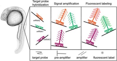 Fig. 1 The RNAscope principle. (Gross-Thebing T, et al., 2014)
Principles of RNAscope ISH
At the core of RNAscope ISH technology lies the innovative probe design and signal amplification approach. The proprietary probe technology features a network of "ZZ" pairs, which enhances the sensitivity and specificity of target RNA detection by forming a branched structure upon hybridization. This branching mechanism enables the amplification of signals at the site of RNA expression, resulting in robust and specific detection of RNA molecules with minimal background noise.
Advantages of RNAscope ISH
The RNAscope ISH technology offers several distinct advantages over traditional RNA in situ hybridization methods.
- Single-molecule sensitivity. RNAscope ISH allows for the precise visualization and quantification of individual RNA molecules within cells and tissues. This single-molecule sensitivity enables the detection of low-abundance transcripts, including rare or novel RNA species, providing invaluable insights into gene expression patterns and regulatory mechanisms.
- High specificity. The technology offers high specificity in detecting target RNA molecules, minimizing non-specific background signals. This high level of specificity ensures accurate and reliable RNA expression data, essential for drawing meaningful conclusions from experimental results.
- Multiplex detection. RNAscope ISH facilitates the concurrent detection of multiple RNA targets within a single tissue section. This capability streamlines the analysis process, enabling researchers to examine the expression patterns of multiple genes simultaneously, thereby enhancing the efficiency and throughput of gene expression analysis.
- Spatial context preservation. By visualizing RNA expression in the context of intact tissue morphology, RNAscope ISH preserves the spatial relationship between cells and their surrounding microenvironment. This feature is particularly valuable in studying developmental processes, tumor heterogeneity, and interactions between different cell types within tissues.
Applications of RNAscope ISH
The versatile nature of RNAscope ISH technology lends itself to a wide range of applications in biomedical research. It has been widely utilized in the fields of developmental biology, cancer research, neuroscience, infectious diseases, and stem cell research, among others.
- Cancer research. RNAscope ISH has been instrumental in characterizing the expression of oncogenes, tumor suppressor genes, and cancer biomarkers within tumor tissues. This technology helps in understanding the cellular heterogeneity within tumors, identifying cancer stem cells, and evaluating the expression of therapeutic targets and drug resistance markers.
- Neuroscience. In neuroscience, RNAscope ISH has proven invaluable for mapping the expression of neurotransmitter receptors, neuropeptides, and neurotrophic factors within the brain and nervous system. It allows for the visualization of gene expression in specific neuronal subtypes, aiding in the study of neural circuitry, synaptic plasticity, and neurodegenerative disorders.
- Infectious diseases. Researchers have utilized RNAscope ISH to study the tropism of pathogenic viruses, localization of viral replication, and host immune responses during viral infections. This technology has provided insights into the cellular targets of viral pathogens and the dynamics of viral spread within infected tissues.
- Stem cell research. RNAscope ISH is employed in the characterization of stem cell populations, differentiation processes, and the expression of lineage-specific markers. It allows for the spatial analysis of gene expression changes during stem cell fate determination and tissue regeneration, contributing to the understanding of stem cell biology and regenerative medicine.
Workflow of RNAscope ISH
The workflow of RNAscope ISH technology encompasses several key steps, including tissue preparation, probe hybridization, signal amplification, and visualization. Tissue sections are prepared and then subjected to target-specific probe hybridization, followed by signal amplification using a sequential enzymatic amplification process. The resulting signal is visualized under a microscope, allowing for the precise localization and quantification of RNA expression within the tissue samples.
Creative Bioarray Relevant Recommendations
| Product/Service Types | Description |
| In Situ Hybridization (ISH) & RNAscope Services | Creative Bioarray offers custom RNA ISH and ACD Bio's RNAscope services. We have extensive experience in the quantification of RNA expression on a cell-by-cell basis across a whole tissue section or within defined regions of interest. |
| ISH Probes | Creative Bioarray offers in situ hybridization probes and specializes in providing high-quality, customizable probes for researchers and laboratories conducting in situ hybridization experiments. |
| Custom Probes | Creative Bioarray is pleased to announce the introduction of customized ISH probes for a range of molecular and cytogenetic applications. |
Reference
- Gross-Thebing T, et al. (2014). "Simultaneous high-resolution detection of multiple transcripts combined with localization of proteins in whole-mount embryos." BMC Biol. 12: 55.