Multicolor FISH (M-FISH) Analysis
- Unlock the Power of Multicolor FISH Analysis for Accurate Genetic Insights
- Service Details
- Features
- Case Studies
- FAQ
- Explore Other Options
Multicolor FISH (M-FISH) assays are used for a precise assessment of complex chromosomal rearrangements. This technique uses all whole-chromosome painting probes in multiplex-FISH and spectral karyotyping. Thus, marker chromosomes, complex chromosomal rearrangements, and all numerical aberrations can be visualized simultaneously in a single hybridization experiment. M-FISH is a kind of versatile tool for precise chromosome separation and for rapid chromosome classification help speeding up the workflow. Apart from that, the analysis of M-FISH images is easy even if aberrations are involved.
Unlike traditional FISH methods, M-FISH allows for the simultaneous visualization of all 24 human chromosomes in a single experiment, providing unparalleled precision and detail. With M-FISH, our expert team can accurately identify chromosomal rearrangements, numerical abnormalities, and subtle structural changes that are often missed by other techniques. This enhanced chromosomal analysis empowers us to deliver more reliable diagnostic insights, enabling better-informed decisions in research and clinical applications.
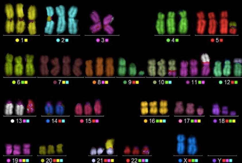
 Fig 1. Representative M-FISH karyotype of K562
cell.
Kamel, Yasser Mostafa, et al. Fluorescence in situ hybridization assays designed for del (7q) detection
uncover more complex rearrangements in myeloid leukemia cell lines. (2014).
Applications of M-FISH:
- Cancer Diagnosis and Prognosis: M-FISH is instrumental in detecting chromosomal rearrangements associated with various cancers, informing treatment decisions and facilitating personalized medicine approaches.
- Genomic Research: This technique allows for the exploration of chromosomal organization and dynamics, contributing to our understanding of gene regulation and chromosomal behavior during cell division.
- Genetic Disorder Diagnosis: M-FISH effectively identifies chromosomal abnormalities linked to genetic disorders, enhancing accuracy in prenatal and postnatal testing.
- Comparative Genomics: M-FISH aids in comparative analyses across species, providing insights into evolutionary relationships and genomic conservation.
Why Choose M-FISH?
Unlike traditional cytogenetic methods, M-FISH offers unparalleled multicolor visualization, allowing for the simultaneous analysis of all chromosomes in a single sample. This leads to improved diagnostic capabilities and more comprehensive genomic insights, making it an essential tool for researchers and healthcare professionals alike.
Key Features

Case Studies
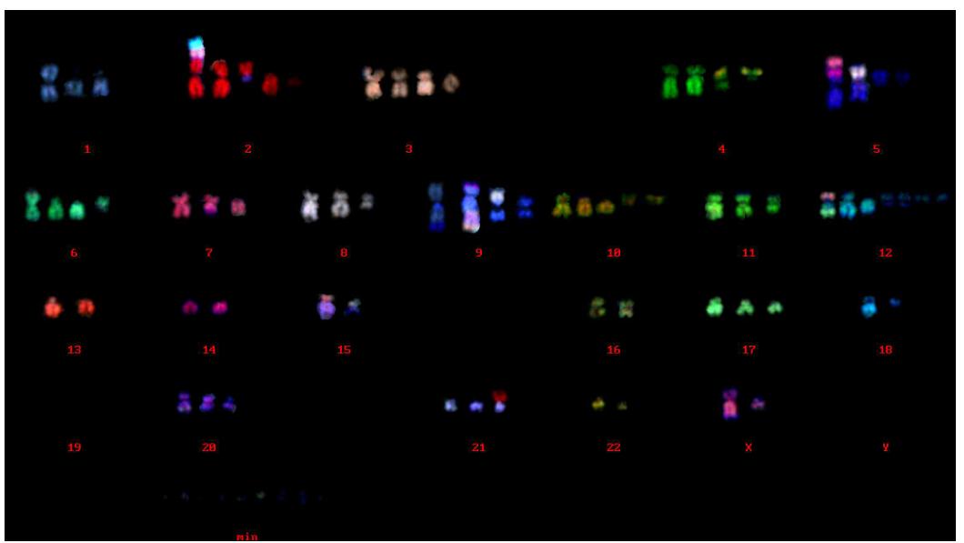
M-FISH analysis of U2OS cell line
Characteristic chromosome rearrangements were found in chromosome X, 2, 5, 9, 12, 15 and 21. The modal number of chromosomes was 74. There were 5-12 of small chromosome fragments in all cells. These fragments were not included in chromosome numbers.
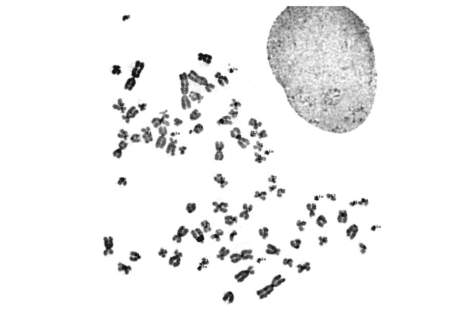
M-FISH karyograms of U2OS cell line

 Fig 2. M-FISH analysis of U2OS cell line
Chromosome number analysis of U2OS cell line
Table 1. Chromosome numbers of U2OS cell line in 50 cells.
| Number of Chromosomes | 66 | 68 | 69 | 70 | 71 | 72 | 73 | 74 | 75 | 78 | 79 |
| U2OS | 2 | 1 | 1 | 2 | 4 | 11 | 7 | 14 | 6 | 1 | 1 |
Results of M-FISH analysis of U2OS cell line
The modal number of chromosomes was 74. There were 5-12 of small chromosome fragments in all cells. They are not included in chromosome numbers. M-FISH karyograms are attached. Characteristic chromosome rearrangements were found in chromosome X, 2, 5, 9, 12, 15 and 21. In the M-FISH karyograms, there were 6-10 of small chromosome fragments in all cells indicated "min" in karyogram. These fragments were undeterminable by M-FISH.
FAQ
1. How does M-FISH differ from traditional FISH?
Unlike traditional FISH methods, which typically identify only one or a few chromosomes at a time, M-FISH provides a comprehensive view of all chromosomes in a single experiment. This multicolor capability enhances the detection of complex chromosomal abnormalities, making it a powerful tool for genomic analysis.
2. What are the primary applications of M-FISH?
M-FISH is widely applied in various fields, including cancer genetics, prenatal diagnostics, developmental biology, and comparative genomics. It is particularly useful for identifying chromosomal abnormalities associated with cancers, genetic disorders, and other chromosomal variations.
3. How is M-FISH performed?
The M-FISH process involves several key steps: sample preparation, hybridization of fluorescent probes to specific chromosomes, imaging using fluorescence microscopy, and subsequent analysis of the captured images. Our experienced team ensures that each step is executed with precision to deliver accurate results.
4. What types of samples can be used for M-FISH analysis?
M-FISH can be performed on a variety of sample types, including cultured cells, tissue samples, and even clinical specimens. Our laboratory is equipped to handle diverse sample preparations tailored to your research or diagnostic needs.
5. How does M-FISH contribute to cancer diagnosis and treatment?
M-FISH plays a crucial role in oncology by identifying chromosomal rearrangements and numerical abnormalities that are often indicative of malignancies. This information aids in diagnosing specific cancer types, determining prognosis, and informing treatment strategies tailored to individual patients.
6. What is the turnaround time for M-FISH results?
The turnaround time for M-FISH analysis can vary depending on the specific project and sample type. Typically, results can be provided within a few weeks. Our team will communicate estimated timelines upfront during the service request process.
7. How can I request M-FISH services?
To request M-FISH analysis or to get more information about our services, please contact us via our website or call our customer service team. We are happy to answer your questions and guide you through the inquiry process.
Quotation and ordering
Our customer service representatives are available 24hr a day! We thank you for choosing Creative Bioarray at your preferred Multicolor FISH (M-FISH) Analysis Service.
References
- Kamel, Yasser Mostafa, et al. "Fluorescence in situ hybridization assays designed for del (7q) detection uncover more complex rearrangements in myeloid leukaemia cell lines. (2014).
- Liehr, Thomas, et al. "Multicolor FISH methods in current clinical diagnostics." Expert review of molecular diagnostics13.3 (2013): 251-255.
- Kearney, L. "Multiplex-FISH (M-FISH): technique, developments and applications." Cytogenetic and genome research 114.3-4 (2006): 189-198.
Explore Other Options