Tumor Cell Culture Guide
Tumor cells occupy the core position in tissue culture. First, tumor cells are relatively easy to culture, and tumor cell lines are the most numerous in currently established cell types. Besides, tumor is the biggest threat to human disease. As a result, tumor cell culture has become an indispensable technology in understanding the mechanism of cancer and developing anticancer drugs. This guide is designed to serve as a basic introduction to tumor cell culture, including media, subculturing, cryopreservation and contamination.
Biological Characteristics of Tumor Cell
Tumor cells are significantly different from normal cells in vivo and in vitro, in terms of morphology, growth value, genetic traits and so on. The difference defined by two tumor cells cultured in vitro are fairly small but not exactly the same. The tumor cells in culture have the following characteristics:
- Immortality
The growth and death of normal cells are strictly controlled. Cells use extracellular signals to regulate the rate and location of division. Generally, apoptosis can be triggered by normal life program. On the contrary, tumor cells provide a mechanism that allows them to bypass the cell cycle checkpoints and inhibit apoptosis. In cancerous cells mitosis can take place continuously, generating an over-abundance of cells to form tumors.
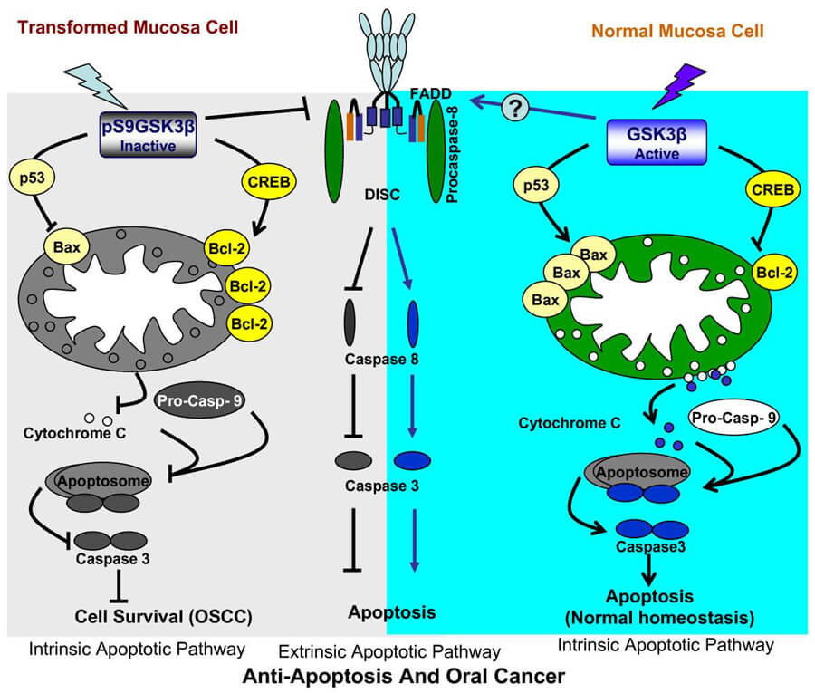 Figure 1. Apoptosis in oral tumor cell and Normal cell. (Rajakishore Mishra, 2010)
- No Contact Inhibition
Normal cells will divide until they are in contact with the neighboring cells, then they will stop growing at that point and form a monolayer cell. While cancer cells typically lose this contact inhibition, causing them to pile up and form tumors (Figure 3).
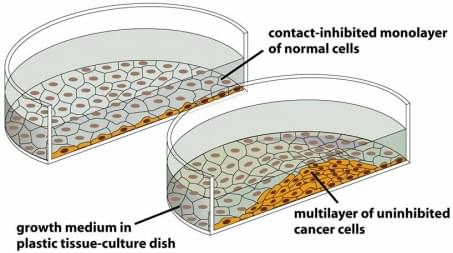 Figure 2. Tumor cell has loosed the contact inhibition.
- Invasiveness
Normal cells normally located at a fixed position until they died. But tumor cells are able to spread into or invade nearby tissues. In addition, some tumor cells can break off and travel to distant places in the body through the blood or the lymph system and form new tumors far from the original tumor.
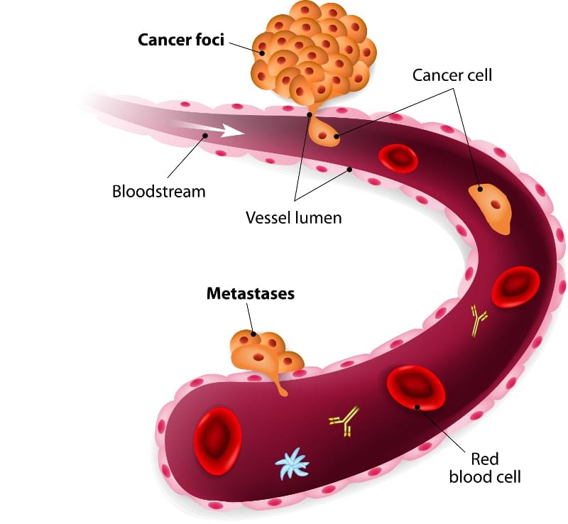 Figure 3. Tumor cells circulate through blood and form a new tumor.
Principles of Tumor Cell Culture
The basics of cell culture are now relatively common, though it was not always so. The prerequisites for cell culture is a well-lit and suitably ventilated laboratory with a laminar flow hood (Class II), CO2 incubator, benchtop centrifuge, microscope, plastic ware (flasks and plates) and a supply of media with or without serum supplements.
Media & Serum
Most numbers of media are a basal saline medium to which various components are added. Based on this saline medium, amino acids, vitamins, metal ions, trace elements, and an energy source (usually provided as glucose) are added to meet cells requirement. Other additives include buffer and phenol red to indicate the acidity of the culture.
Table 1. Common media list.
| Medium | Application |
Basal Medium Eagle (BME) | Diploid and primary cell cultures, such as HeLa, L-cells and primary mammalian fibroblasts |
| Minimum Essential Medium (MEM) | Suspension and adherent mammalian cells |
| DMEM | Preserve and maintain the growth of most mammalian cells |
| MCDB | Human microvascular endothelial cells |
| Most mammalian and hybridoma cells, such as human myeloma, mouse hybridoma, human leukocytes, B and T lymphocytes |
Serum is often added to the media described above to replace many requirements which are missing. The concentration of components present is often variable from 5-20%, depending on the cell type, and the practice of using newborn or fetal bovine sera has become common. It is better to stick with one supplier found to work well with a particular cell line due to variation exists in batch to batch. Serum can be divided into aliquots and frozen at −20°C.
There are some cell types that can't culture in a media contain sera, such as human microvascular endothelial cells. Add specific supplements or increase the concentration of various components of existing basal media are both available to meet special requirements.
Subculturing
Subculturing is a process that a new cell or microbiological culture made by transferring some or all cells from a previous culture to fresh growth medium. Different cell types have particular subculturing requirements, a preferred range of densities and a good condition for optimal growth. So more details please refer to the specific technical bulletins on our website.
Cryopreservation
In order to store cells effectively and then recover a high enough proportion for quality research, storage conditions must be monitored on a permanent basis and excellent records kept.
Cellular damage induced by freezing and thawing is generally believed to be caused by intracellular ice crystals and osmotic effects. The addition of a cry protective agent, such as dimethyl sulfoxide (DMSO) or glycerol, and the selection of suitable freezing and thawing rates minimizes cellular injury. While short-term storage of cell lines using mechanical freezers (-75°C) is possible, storage in liquid nitrogen (-196°C) or its vapour (to -135°C) is much preferred. The use of liquid nitrogen refrigerators is advantageous not only because of the lower temperatures and, consequently, almost infinite storage times possible, but also due to the total absence of risk of mechanical failures and the prolonged holding times now available.
Contamination
Cell culture should be stain sterility, and regular sterility testing is necessary. Although cell cultures may be produced within endogenous (mainly viral) infections, most infections and contamination are acquired. Here are some common culture contaminations but not limited:
- Bacterial
Bacteria are a large and ubiquitous group of unicellular microorganisms. Because of their ubiquity, size, and fast growth rates, bacteria are one of the most commonly encountered biological contaminants in cell culture. If present, it is easily detected by visual inspection of the phenol red will turn yellow and the cells will be replaced by colonies of bacteria under the inverted microscope. It is common practice to add antibiotics to cultures and this can mask low level infection for a considerable time.
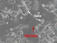 Figure 4. Cell contamination by bacteria.
- Mold
Molds are eukaryotic microorganisms in the kingdom of Fungi. Different from bacteria contamination, the pH of the culture will remain stable in the initial stages of contamination, then rapidly increases as the culture become more heavily infected and becomes turbid. Under microscopy, the mycelia usually appeared. Generally, add antibiotics to cultures and it will be able to decrease the level of infection to an extent.
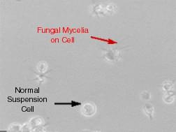 Figure 5. Cell contamination by Mold.
- Yeasts
Yeasts are unicellular eukaryotic microorganisms, similar to mold contamination, cultures contaminated with yeasts become turbid, especially if the contamination is in an advanced stage. The pH of the culture contaminated by yeasts may not change too much until the contamination becomes heavy, at which stage the pH usually increases. Under microscopy, it is obvious that yeast appears as individual ovoid or spherical particles and even may bud off smaller particles. Add antibiotics to cultures did not work very well. Most laboratories usually dispose of infected cultures and start again.
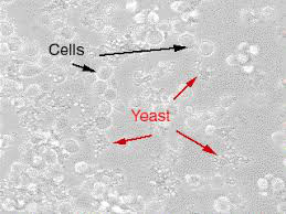 Figure 6. Cell contamination by yeast.
- Mycoplasma
Mycoplasma maybe the most common and most missed infection in cell culture laboratories, and their effects are insidious. The indicators that there might be a problem include reduced growth rate, morphological changes, chromosomal aberration, and altered metabolism. There are several ways of testing for Mycoplasma and many manufacturers provide kits.
- Virus
Some cell lines contain viruses including integrated into their genome, or as endogenous non-lethal infections. In many cases, these are not regarded as infections, and viral transformation of cells is a time-honored method of producing continuous cell lines. However, using virally infected cell cultures can present a serious health hazard to the laboratory personnel, especially if human or primate cells are cultured in the laboratory.
Getting Started with Creative Bioarray
Creative Bioarray provides multiple kinds of tumor cells from human or animal which will be delivered by the cold chain transport. Each cell product from Creative Bioarray has a product sheet that contains detailed information for handling the cells such as the number of cells and the recommended subcultivation ratio. And all these product sheets can be found at the website or you can request a copy via sending an e-mail to our technology platform.
Creative Bioarray is dedicated to providing best cell products and services to accelerate the achievement of your research goals. We also offer customized service to meet all your special requirements. We will be your best and most reliable partner, looking forward to cooperating with you!
References
- Mishra R. Glycogen synthase kinase 3 beta: can it be a target for oral cancer. Molecular Cancer. 2010, 9:144.
- Cree I A. Cancer Cell Culture[M]. Springer Berlin, 2011.
- Mather J P, Barnes D W. Animal cell culture methods[M]. Academic Press, 1998.