Exploring Cell Dynamics: Migration, Invasion, Adhesion, Angiogenesis, and EMT Assays
Cell Migration and Invasion
Cell migration is how cells transfer and orient themselves from one place to another by their own locomotion after receiving a stimulus. It is also involved in a number of physiological and pathological mechanisms, including embryogenesis, inflammatory response, wound healing, immune response, and tumor invasion and dissemination.
Cell migration can be delineated into four major steps:
(1) Extension of lamellipodia at the leading edge of the cell;
(2) Formation of new adhesions between the leading edge of the cell and the extracellular matrix (ECM);
(3) Contraction of the cell body;
(4) Detachment of the trailing edge from the surrounding matrix, enabling forward movement.
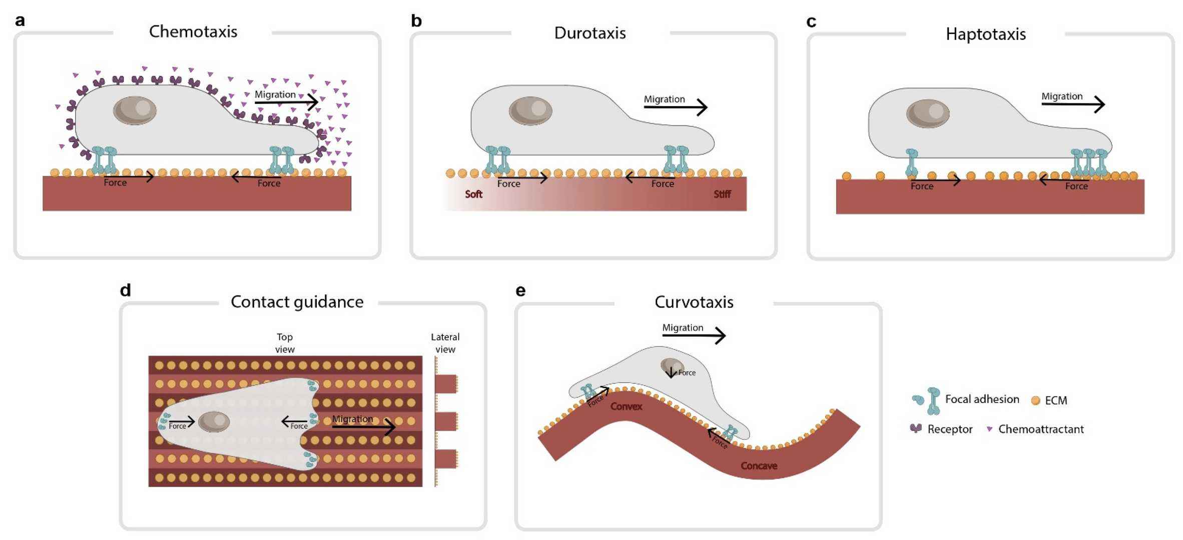 Fig. 1. Common types of directed cell migration (Fortunato IC, Sunyer R. 2022).
A specific kind of cell migration is cell invasion, when invading cells release proteases to break down the ECM or BME to make their way into blood vessels and lymphatic vessels (common in malignant tumors).
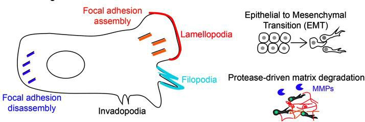 Fig. 2. The metastatic cascade and cancer cell migration (Payne SL, Levin M, et al., 2019).
Cell migration and invasion assay
- Scratch Assay: The Scratch Assay, also known as the Wound Healing Assay, is one of the earliest developed methods for detecting in vitro cell migration. The basic steps include creating a "wound" on a confluent cell monolayer, capturing images at the onset of and during the cell migration process to close the wound, and comparing these images to determine the rate of cell migration.
- Transwell Assay: The Transwell Assay is an experimental technique that can effectively assess both cell migration and invasion capabilities. Cells are placed in the upper chamber of the Transwell insert, and the lower chamber contains chemoattractants that attract cell migration. This setup allows cells to migrate through the pores of a polycarbonate membrane from the upper chamber to the lower chamber, simulating their response and movement in vivo to chemical signals.
- RTCA Technology: Real-Time Cellular Analysis (RTCA) is a method that uses advanced methods to integrate microelectronic cell sensor chips into the bottom of the detection plate, allowing for real-time, dynamic, and quantitative tracking of cell migration and invasion dynamics.
- 3D Cell Invasion Assay: By simulating the in vivo environment, cells are embedded within a three-dimensional matrix to study their migration and invasion behaviors. This method more closely resembles actual in vivo conditions.
Cell Adhesion
Cell adhesion refers to the attachment and interaction between cells or cells with their matrix. This is a critical part of many biological processes, such as tissue formation, cell communication, tissue repair, immune responses, and cancer growth. Cell adhesion assays are used to assess the effects of growth factors, chemicals, drugs and genetic engineering on cell adhesion.
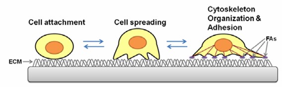 Fig. 3. The process of cell adhesion (Xi J, et al., 2013).
- Cell Counting Method: Cells are seeded on a surface of a substrate or culture plate. After growing, the unattached cells are taken out, and the adhered cells are fixed and stained with fluorescent dyes or dyes such as crystal violet and MTT. This is then evaluated with a fluorescence microscope or plate reader to check the adhesion state.
- Microfluidic Technology: Microfluidic chips mimic physiological fluid environments in vivo, enabling precise cell and fluid control. The technology allows for a clear view of cell adhesion to substrates in flow.
- Atomic Force Microscopy (AFM): AFM detects interactions by measuring the bend of a small cantilever when its end passes through a cell. It could measure the force of adhesion between cells and substrates.
- Cell Adhesion Force Test- Centrifugation Process: Cells are spun by centrifugal force and released from the substrate. The percentage of cells that resist this force and remain stuck is calculated to measure the strength of adhesion.
Angiogenesis
Angiogenesis, the creation of new blood vessels from existing capillaries or post-capillary venules, is central to crucial physiological processes such as organ growth, embryogenesis and wound healing. Angiogenesis involves six stages:
(1) Endothelial cell activation,
(2) signal transduction,
(3) matrix degradation,
(4) cell invasion,
(5) lumen formation,
(6) vessel maturation.
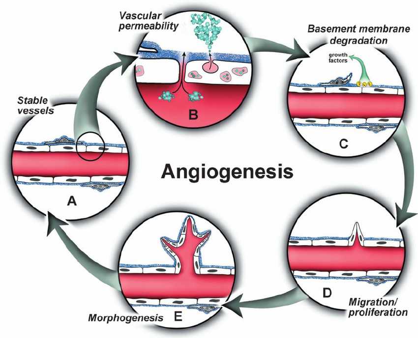 Fig. 4. The angiogenic cascade (Bryan BA, D'Amore PA, 2007).
In vitro angiogenesis assay
This is one of the most traditional methods to study angiogenesis in vitro that usually involves endothelial cells such as human umbilical vein endothelial cells (HUVECs) that have a tendency to multiply and move rapidly. When cultured on basement membrane extracts (BME), the cells are three-dimensional capillary tubular structures and therefore can be manipulated to identify angiogenesis inhibitors and activators.
Epithelial-Mesenchymal Transition
Epithelial-Mesenchymal Transition (EMT) is an important event that takes place in development, wound healing and the development of cancers as epithelial cells become mesenchymal. This change promotes better cell movement, decreases intracellular adhesion and increases secretion of extracellular matrix molecules that lead to the transition to fibroblast-like shape. EMT is essential for embryogenesis, organogenesis, wound healing and cancer development. In cancer, EMT allows cells to gain invasive and metastatic properties, which help spread cancer cells, and potentially present new targets for cancer treatment.
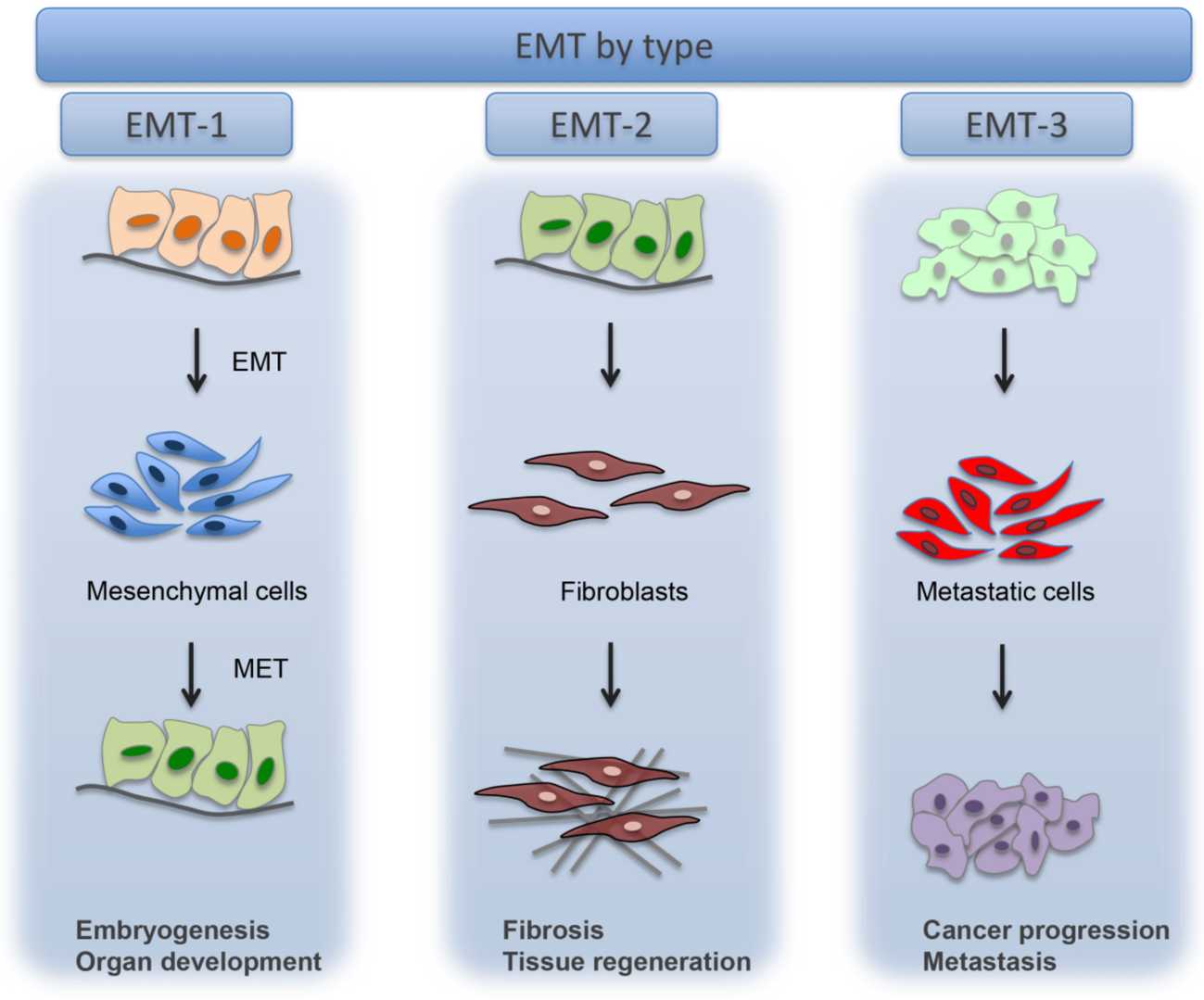 Fig. 5. Types of EMT (Marconi GD, Fonticoli L, et al., 2021).
Detection of EMT markers:
The expression changes of EMT marker proteins (E-cadherin, N-cadherin, Vimentin) can be assessed by Western blot and real-time quantitative PCR, or directly observed by immunofluorescence labelling of EMT markers.
Detection of EMT phenotypic changes:
The morphology of cells changing from epithelial to mesenchymal during EMT is easy to monitor through direct observation.
In addition, EMT causes cell-migration and invasion activity changes, as can be tested by scratch assays, Transwell invasion assays and so on.
Creative Bioarray Relevant Recommendations
| Products & Services | Description |
| Cell Migration and Invasion Assay Services | Creative Bioarray provides cell migration and Invasion Assay Services to all of our customers. Two methods are available, which are Cell culture wound closure assay and Transwell cell migration and invasion assay. |
| 3D Invasion Assay | Creative-Bioarray offers rapid, automatable 3D invasion system that enables highly reproducible experimental conditions and high throughput analysis will give our customs the best results to identify novel therapeutic agents. |
| Cell Angiogenesis Assays | Creative Bioarray provides a series of in vitro and in vivo angiogenesis assays to support the development of angiogenic or inhibitory compounds. |
| Cellular Phosphorylation Assays | With 50 Kinase cell lines available, Creative Bioarray provides several different cellular phosphorylation assays to accelerate your research of kinase functional activity, dimerization, characterization, and inhibitor screening. We provide technical methods of ELISA or Western Blot based on the customer's requirements. |
References
- Fortunato IC, Sunyer R. The Forces behind Directed Cell Migration. Biophysica. 2022. 2(4):548-563.
- Payne SL, Levin M, et al. Bioelectric Control of Metastasis in Solid Tumors. Bioelectricity. 2019. 1(3):114-130.
- Xi J, et al. Quartz crystal microbalance in cell biology studies. J Biochip Tissue chip S, 2013. pp: 2153-0777.
- Bryan BA, D'Amore PA. What tangled webs they weave: Rho-GTPase control of angiogenesis. Cell Mol Life Sci. 2007. 64(16):2053-65.
- Marconi GD, Fonticoli L, et al. Epithelial-Mesenchymal Transition (EMT): The Type-2 EMT in Wound Healing, Tissue Regeneration and Organ Fibrosis. Cells. 2021. 10(7):1587.