In Situ Hybridization (ISH) and RNAscope Services
- Service Details
- Workflow
- Features
- Applications
- FAQ
In situ hybridization (ISH) is a technique that enables researchers to directly visualize and localize specific nucleic acid sequences (DNA or RNA) without compromising the structural integrity of cells or tissues. With continuous technological advancements, ISH has evolved from traditional DNA ISH to more refined RNA ISH, and more recently, to the revolutionary RNA detection service—RNAscope—introduced by ADC Company. The advanced capabilities of these technologies deliver extraordinary precision and insights for gene expression research and disease diagnostics as well as drug development applications.
At Creative Bioarray, we offer customized RNA ISH and RNAscope services, delivering comprehensive solutions ranging from routine detection to single-molecule-level analysis. Our services empower breakthroughs in tissue microenvironment exploration, single-cell heterogeneity studies, and biomarker discovery.
Our In Situ Hybridization Services Methods
We provide comprehensive ISH testing solutions to advance gene expression analysis in research and drug discovery. Here are our core testing methods:
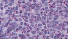
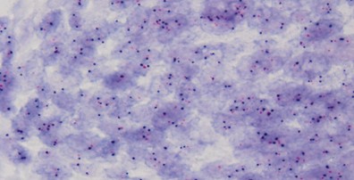
RNAscope
RNAscope is a proprietary, ultra-sensitive assay that enables single-cell resolution detection of RNA targets within intact tissue architecture, preserving spatial and morphological context. Its double-Z probe design ensures exceptional specificity for low-abundance transcripts, such as long non-coding RNAs (lncRNAs) or viral RNA.
Basescope
BaseScope specializes in detecting ultra-short RNA sequences (as few as 50 nucleotides) with single-molecule precision. This technology uniquely resolves highly homologous sequences, including splice variants, single-nucleotide polymorphisms (SNPs), or strain-specific viral RNAs.
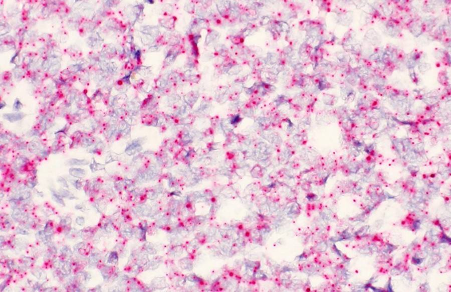
CISH
CISH employs enzyme-linked chromogenic probes to generate stable, brightfield-compatible signals in formalin-fixed, paraffin-embedded (FFPE) tissues. This cost-effective method is ideal for DNA/RNA localization and biomarker validation (e.g., HER2/neu, EBER).
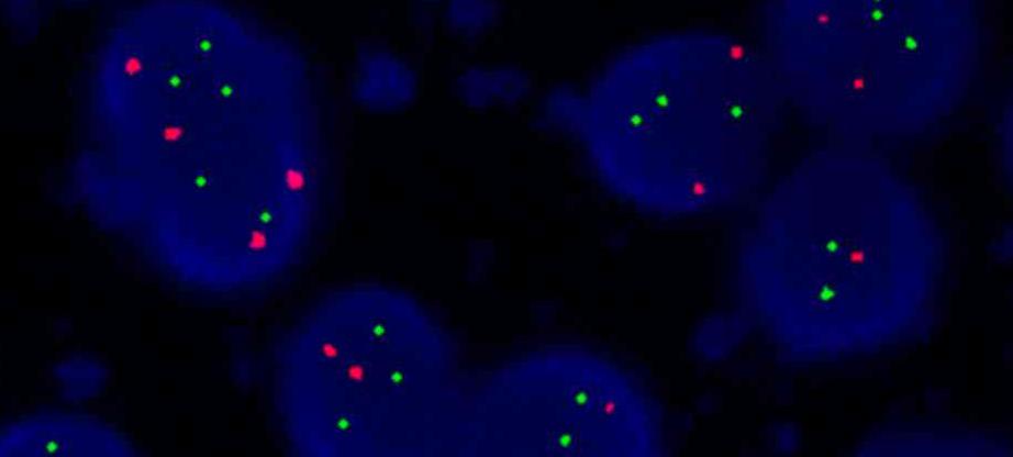
FISH
FISH utilizes fluorophore-labeled probes for multiplexed, high-resolution spatial analysis of nucleic acids. It supports dynamic RNA/DNA co-localization, 3D-tissue imaging, and integration with protein marker detection.
RNA Types:
- mRNA;
- non-coding RNAs (miRNA, lncRNA);
- viral RNA (e.g., HPV, HIV);
- engineered transgene RNA…
Sample Types:
- FFPE tissues;
- fresh-frozen tissues;
- CTCs;
- cell line;
- blood…
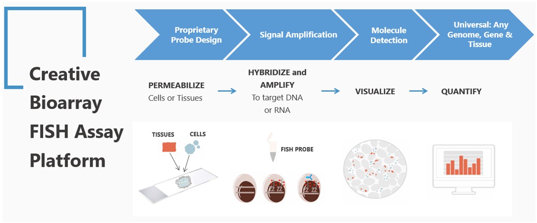
Workflow
1
Pretreatment
- FFPE samples: Deparaffinization, rehydration, and protease digestion to expose target RNA.
- Frozen sections: Fixation followed by permeabilization to enhance probe penetration.
2
Probe preparation
Design and label target-specific probes based on RNA sequences; validate binding efficiency and specificity via pre-experiments.
3
Hybridization and signal amplification
Hybridize probes with target RNA under controlled conditions; amplify signals using specialized techniques (e.g., enzymatic or Tyramide Signal Amplification).
4
Detection and analysis
- Capture bright-field or fluorescence images using microscopy.
- Analyze data with AI-powered software to quantify expression intensity, positive cell ratios, and spatial distribution heatmaps.
5
Report delivery
Provide high-resolution images, quantitative charts, and spatial visualization results.
Key Features

High Sensitivity & Specificity
RNAscope technology enables single-molecule-level detection, ensuring accurate identification of low-abundance targets.

Customized Solutions
Tailored probe design, sample processing, and protocol optimization to meet specific research needs.

Comprehensive Data Analysis
High-quality image analysis and quantitative data for in-depth interpretation.

Rapid Turnaround
Streamlined workflows ensure efficient service delivery.
What Can You Do with Our ISH and RNAscope Services
Basic Research: Gene expression profiling, cell differentiation studies, developmental biology, and disease mechanism exploration.
Disease Diagnosis: Detection of genetic disorders, tumor biomarkers, viral infections (e.g., HPV, HIV), and drug target validation.
Drug Development: Evaluation of gene therapy vector expression, CAR-T cell monitoring, and drug target screening.
FAQ
Q1: Are there any restrictions on sample types?
We support diverse sample types, including FFPE tissues, frozen sections, and cell samples. Contact our technical team for pre-evaluation if uncertain.
Q2: How should tissue samples be prepared?
FFPE samples require unstained consecutive sections (4-5 μm thickness) to prevent RNA degradation. Frozen samples must be preserved under RNase-free conditions.
Q3: What distinguishes RNA ISH from RNAscope?
RNA ISH is a traditional method for routine RNA detection, while RNAscope offers superior sensitivity for low-abundance targets with minimal background interference.
Q4: What is the typical turnaround time?
Standard projects take 2-3 weeks, depending on sample type and assay complexity.
Q5: Do you offer custom probe design?
Yes, species-specific probes can be designed upon request.
References
- Duderstadt EL, Sanders MA, et al. A Method to Pre-Screen Rat Mammary Gland Whole-Mounts Prior To RNAscope. J Mammary Gland Biol Neoplasia. 2021. 26(2):113-120.
- Hammer PM, Wang A, et al. Detection of FOXL2 C134W Mutation Status by a Novel BaseScope In Situ Hybridization Assay is Highly Sensitive and Specific for Adult Granulosa Cell Tumors. Mod Pathol. 2023. 36(11):100318.
- Hu YC, Bandla S, et al. HER2 amplification, overexpression and score criteria in esophageal adenocarcinoma. Modern Pathology. 2011. 24(7):899-907.
- Cao P, Yu Y, et al. Fluorescence in situ hybridization comparison of the prognostic factors in adult and pediatric acute lymphoblastic leukemia: A retrospective analysis of 282 cases. Exp Ther Med. 2018 Dec;16(6):4674-4684.
Explore Other Options