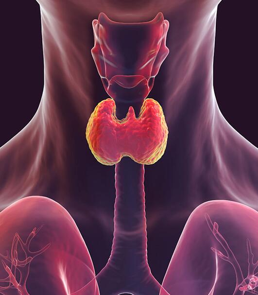
Thyroid Tumor Cells
Thyroid tumors encompass a wide spectrum of malignancies, from well-differentiated follicular and papillary carcinomas to aggressive anaplastic and medullary types. These tumors are characterized by distinct genetic alterations and varying degrees of cellular differentiation, posing significant challenges for both diagnosis and treatment.
Our thyroid tumor cell line collection offers a diverse and authentic set of in vitro models derived directly from human patient specimens. These models reflect the biological complexity and heterogeneity of thyroid cancers, providing essential tools for investigating tumor progression, exploring targeted therapeutic strategies, and studying mechanisms of treatment resistance.
Authentic Genetically Diverse Clinically Relevant Rigorously Tested
Key Features & Expertise
Our thyroid tumor cell lines stand out for the following features
- Covers a broad range of thyroid malignancies, including follicular, papillary, and anaplastic subtypes
- Includes models derived from primary tumors, lymph node metastases, and pleural effusions
- Represents various patient demographics and clinical presentations
- Genomic and phenotypic profiling available for key alterations
- Documented growth characteristics suitable for reproducible in vitro studies
- Consistent behavior across passages for reliable experimental outcomes
- STR-authenticated to ensure cell line identity and purity
- Mycoplasma-free and routinely screened for contamination
- Stable supply with comprehensive technical support
FAQ
What types of thyroid cancer are represented in your cell line collection?
How do I choose the right thyroid cancer cell line for my research?
- Tumor subtype and differentiation status (e.g., follicular vs. anaplastic)
- Genetic background and known mutations
- Growth behavior and culture requirements
- Whether you need a model for drug screening, metastasis studies, or mechanistic research
Are your thyroid tumor cell lines authenticated?
Can these cell lines be used for drug screening and resistance studies?
How stable are thyroid tumor cell lines during long-term culture?
Do you provide information on the source and history of each cell line?
How are the cell lines shipped and stored?
Filters Clear all filters
Species
- Cat (1)
- Human (989)
- Mouse (5)
- Rat (1)
Source
- Abdomen Metastasis (2)
- Adrenal Gland (7)
- Adrenal Gland Metastasis (2)
- Ascites (23)
- Ascites Metastasis (32)
- Bile Duct (3)
- Bladder (12)
- Blood (120)
- Bone (21)
- Bone Marrow (43)
- Bone Marrow Metastasis (18)
- Bone Metastasis (6)
- Brain (31)
- Brain Metastasis (6)
- Breast (8)
- Bronchus (1)
- Cecum (3)
- Cerebrospinal Fluid (1)
- Cerebrospinal Fluid Metastasis (1)
- Cervix (32)
- Colon (83)
- Cornea (3)
- Cutaneous Metastasis (1)
- Dermis (1)
- Duodenum (1)
- Endometrium (17)
- Esophagus (44)
- Eye (12)
- Eye Socket (5)
- Fetus (1)
- Foreskin (4)
- Gallbladder (1)
- Gingiva (2)
- Globe (2)
- Groin (1)
- Hypodermis Metastasis (5)
- Intestine (84)
- kidney (1)
- Kidney (9)
- Liver (13)
- Liver Metastasis (17)
- Lung (42)
- Lung Metastasis (8)
- Lymph Node (5)
- Lymph Node Metastasis (56)
- Muscle (4)
- Muscle Metastasis (2)
- Nose (2)
- Omentum Metastasis (2)
- Oral Cavity (10)
- Ovary (13)
- Ovary Metastasis (2)
- Pancreas (10)
- Pelvic Wall Metastasis (1)
- Pelvis (1)
- Perianal Space Metastasis (1)
- Pericardial Effusion (1)
- Pericardial Effusion Metastasis (1)
- Perineus (1)
- Peripheral Blood (119)
- Peritoneal Effusion (2)
- Peritoneum (1)
- Peritoneum Metastasis (1)
- Pharynx (3)
- Pleural Effusion (54)
- Pleural Effusion Metastasis (44)
- Prostate (4)
- Rectum (13)
- Renal Pelvis (1)
- Retroperitoneal Space (2)
- Salivary Gland (2)
- Skeletal Muscle (1)
- Skin (22)
- Skin Metastasis (3)
- Small Intestine (1)
- Small Intestine Metastasis (1)
- Soft Tissue Metastasis (1)
- Stomach (4)
- Testis (9)
- Thoracic Cavity Metastasis (6)
- Thyroid Gland (15)
- Thyroid Gland Metastasis (1)
- Tongue (5)
- Umbilical Cord (1)
- Umbilical Cord Blood (1)
- Urachus (1)
- Ureter (1)
- Uterus (53)
- Uvea (2)
- Vagina (2)
- Vulva (1)
Disease
- Acute Biphenotypic Leukemia (1)
- Acute Erythroid Leukemia (4)
- Acute Megakaryoblastic Leukemia (4)
- Acute Monocytic Leukemia (9)
- Acute Myeloid Leukemia (25)
- Acute Promyelocytic Leukemia (2)
- Adrenal Gland Neuroblastoma (11)
- Adult B Acute Lymphoblastic leukemia (1)
- Adult B Acute Lymphoblastic Leukemia (6)
- Adult T Acute Lymphoblastic Leukemia (6)
- Adult T Lymphoblastic Lymphoma (2)
- Adult T-Cell Leukemia/Lymphoma (1)
- Alveolar Rhabdomyosarcoma (4)
- Alveolar Ridge Squamous Cell Carcinoma (1)
- Amelanotic Melanoma (3)
- Ampulla of Vater Adenocarcinoma (1)
- Ampulla of Vater Adenosquamous Carcinoma (3)
- Anaplastic Astrocytoma (3)
- Anaplastic Large Cell Lymphoma (7)
- Askin Tumor (1)
- Astrocytoma (5)
- B Acute Lymphoblastic Leukemia (2)
- B-Cell Non-Hodgkin Lymphoma (5)
- Bare Lymphocyte Syndrome Type 2 (1)
- Barrett Adenocarcinoma (2)
- Benign Prostatic Hyperplasia (1)
- Bladder Carcinoma (12)
- Bladder Squamous Cell Carcinoma (1)
- Breast Adenocarcinoma (1)
- Breast Carcinoma (9)
- Breast Ductal Carcinoma (2)
- Burkitt Lymphoma (17)
- Canavan Disease (1)
- Cecum Adenocarcinoma (3)
- Central Nervous System Lymphoma (2)
- Cervical Adenocarcinoma (2)
- Cervical Adenosquamous Carcinoma (2)
- Cervical Small Cell Carcinoma (1)
- Cervical Squamous Cell Carcinoma (2)
- Childhood B Acute Lymphoblastic Leukemia (13)
- Childhood T Acute Lymphoblastic Leukemia (16)
- Childhood T Lymphoblastic Lymphoma (1)
- Cholangiocarcinoma (2)
- Chronic Eosinophilic Leukemia (1)
- Chronic Lymphocytic Leukemia (2)
- Chronic Myeloid Leukemia (23)
- Clear Cell Renal Cell Carcinoma (2)
- Colon Adenocarcinoma (53)
- Colon Carcinoma (33)
- Colorectal Adenocarcinoma (1)
- Colorectal Carcinoma (1)
- Congenital Pure Red Cell Aplasia (1)
- Cutaneous Melanoma (10)
- Dedifferentiated Chondrosarcoma (1)
- Desmoplastic Melanoma (1)
- Diffuse Large B-Cell Lymphoma (28)
- Down Syndrome (2)
- EBV-Related Burkitt Lymphoma (12)
- Embryonal Carcinoma (3)
- Embryonal Rhabdomyosarcoma (3)
- Endometrial Adenocarcinoma (13)
- Endometrial Adenosquamous Carcinoma (2)
- Endometrial Carcinoma (2)
- Endometrioid Stromal Sarcoma (1)
- Epithelioid Hemangioendothelioma (1)
- Epithelioid Sarcoma (3)
- Esophageal Adenocarcinoma (6)
- Esophageal Squamous Cell Carcinoma (41)
- Essential Thrombocythemia (1)
- Ewing Sarcoma (2)
- Extraskeletal Myxoid Chondrosarcoma (1)
- Fanconi Anemia (1)
- Fibrosarcoma (1)
- Follicular Lymphoma (2)
- Gallbladder Carcinoma (2)
- Gallbladder Undifferentiated Carcinoma (2)
- Gastric Adenocarcinoma (6)
- Gastric Adenosquamous Carcinoma (1)
- Gastric Carcinoma (5)
- Gastric Choriocarcinoma (1)
- Gastric Fundus Carcinoma (1)
- Gastric Signet Ring Cell Adenocarcinoma (1)
- Gastric Small Cell Carcinoma (2)
- Gastric Tubular Adenocarcinoma (5)
- Gastroesophageal Junction Adenocarcinoma (1)
- Gestational Choriocarcinoma (1)
- Gingival Squamous Cell Carcinoma (2)
- Glioblastoma (18)
- Gliosarcoma (1)
- Hairy Cell Leukemia (1)
- Hepatoblastoma (2)
- Hepatocellular Carcinoma (6)
- Hepatosplenic T-Cell Lymphoma (2)
- Hereditary Thyroid Gland Medullary Carcinoma (1)
- High Grade B-Cell Lymphoma (1)
- High Grade Ovarian Serous Adenocarcinoma (8)
- Hodgkin Lymphoma (9)
- Hypopharyngeal Squamous Cell Carcinoma (2)
- Infectious Mononucleosis (1)
- Intrahepatic Cholangiocarcinoma (6)
- Invasive Breast Carcinoma of No Special Type (12)
- Kidney Neoplasm (1)
- Kidney Rhabdoid Tumor (1)
- Krukenberg Tumor (1)
- Liposarcoma (1)
- Lung Adenocarcinoma (17)
- Lung Giant Cell Carcinoma (8)
- Lung Large Cell Carcinoma (9)
- Lung Mucoepidermoid Carcinoma (1)
- Lung Non-Small Cell Carcinoma (2)
- Lung Small Cell Carcinoma (25)
- Lung Squamous Cell Carcinoma (9)
- Lymphoblastic Lymphoma (1)
- Malignant Peripheral Nerve Sheath Tumor (1)
- Mantle Cell Lymphoma (5)
- Mature Gastric Teratoma (1)
- Maxillary Sinus Squamous Cell Carcinoma (1)
- Medulloblastoma (3)
- Melanoma (24)
- Meningioma (2)
- Minimally Invasive Lung Adenocarcinoma (1)
- Monophasic Synovial Sarcoma (1)
- Mouse Intestinal Tract Neuroendocrine Adenoma (1)
- Mouse Mammary Gland Malignant Neoplasm (2)
- Mouse Plasmacytoma (1)
- Mycosis Fungoides (1)
- Myelodysplastic Syndrome (1)
- Myxofibrosarcoma (1)
- Natural Killer Cell Lymphoblastic Leukemia/Lymphoma (2)
- Neuroblastoma (26)
- Oral Cavity Squamous Cell Carcinoma (15)
- Osteoid Osteoma (1)
- Osteosarcoma (15)
- Ovarian Carcinoma (1)
- Ovarian Clear Cell Adenocarcinoma (1)
- Ovarian Endometrioid Adenocarcinoma (4)
- Ovarian Granulosa Cell Tumor (1)
- Ovarian Mucinous Adenocarcinoma (2)
- Ovarian Serous Adenocarcinoma (2)
- Ovarian Serous Cystadenocarcinoma (2)
- Ovarian Small Cell Carcinoma (1)
- Pancreatic Adenocarcinoma (13)
- Pancreatic Carcinoma (5)
- Pancreatic Ductal Adenocarcinoma (12)
- Papillomavirus-Independent Cervical Squamous Cell Carcinoma (1)
- Papillomavirus-Related Cervical Adenocarcinoma (7)
- Papillomavirus-Related Cervical Squamous Cell Carcinoma (4)
- Papillomavirus-Related Endocervical Adenocarcinoma (16)
- Paroxysmal Nocturnal Hemoglobinuria (3)
- Pharyngeal Squamous Cell Carcinoma (1)
- Plasma Cell Myeloma (15)
- Pleural Epithelioid Mesothelioma (5)
- Pleural Sarcomatoid Mesothelioma (2)
- Poorly Differentiated Thyroid Gland Carcinoma (1)
- Primary Cutaneous T-Cell Non-Hodgkin Lymphoma (1)
- Primary Effusion Lymphoma (7)
- Primitive Neuroectodermal Tumor (1)
- Prostate carcinoma (1)
- Prostate Carcinoma (9)
- Rectal Adenocarcinoma (13)
- Rectosigmoid Adenocarcinoma (1)
- Recurrent Bladder Carcinoma (1)
- Renal Cell Carcinoma (7)
- Renal Pelvis Urothelial Carcinoma (1)
- Retinoblastoma (11)
- Sacral Chordoma (1)
- Sacrococcygeal Teratoma (1)
- Salivary Gland Squamous Cell Carcinoma (1)
- Sezary Syndrome (1)
- Shwachman-Diamond Syndrome (1)
- Skin Squamous Cell Carcinoma (2)
- Splenic Marginal Zone Lymphoma (1)
- Testicular Embryonal Carcinoma (8)
- Testicular Teratoma (2)
- Testicular Yolk Sac Tumor (1)
- Thyroid Gland Anaplastic Carcinoma (10)
- Thyroid Gland Follicular Carcinoma (4)
- Thyroid Gland Papillary Carcinoma (3)
- Thyroid Gland Sarcoma (1)
- Thyroid Gland Squamous Cell Carcinoma (2)
- Tongue Adenosquamous Carcinoma (1)
- Tongue Squamous Cell Carcinoma (6)
- Type I Endometrial Adenocarcinoma (1)
- Ureter Urothelial Carcinoma (1)
- Uterine Carcinosarcoma (2)
- Uterine Corpus Leiomyosarcoma (1)
- Uterine Corpus Sarcoma (2)
- Uveal Melanoma (2)
- Vaginal Melanoma (2)
- Vulvar Melanoma (1)
- Vulvar Squamous Cell Carcinoma (1)

Description: FTC-133 was isolated from a lymph node metastasis of a follicular thyroid carcinoma. The morphology ...

Description: Species: human male 42 years old; Tissue: thyroid; Tumor: carcinoma, follicular; Derived from: lung ...
Description: Species: human; Tissue: thyroid; Tumor: carcinoma, follicular

Description: Established from the primary tumor of a 67-year-old woman with primary thyroid carcinoma ...
Description: Established from the sarcoma of the thyroid gland from a 47-year-old woman in 1985

Description: Established from the tumor tissue of a 76-year-old woman with metastasizing papillary thyroid ...

Description: established from the lymph node metastasis of a 63-year-old woman with anaplastic papillary thyroid ...

Description: Established from the sternal metastasis of follicular thyroid carcinoma removed from a 70-year-old ...

Description: Established from the thyroid gland (right lobe) of a 70-year-old woman with thyroid anaplastic ...

Description: ML-1 is one of three celll lines isolated in 1978 from the peripheral blood of a 24 year old male ...

Description: Established from the malignant tumor of a 49-year-old woman with well-differentiated oxyphilic ...

Description: Established in 1996 from the tumor tissue of a 57-year-old Caucasian man with local recurrence of a ...

Description: Established from cancer cells disseminated in the pleural fluid of a 44-year-old woman with ...

Description: Established from the primary tumor of a 78-year-old woman with thyroid carcinoma (undifferentiated ...
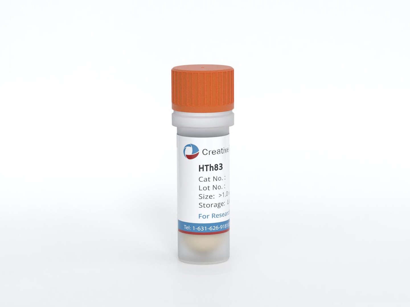
Description: established from the thyroid gland from a 66-year-old male patient with thyroid gland anaplastic ...

Description: G-CSF producing thyroidal squamous carcinoma.

Description: Serum-free, protein-free cultured HOTHC cells.

Description: Thyroid tumor. TAF, G-CSF, colloid producing.