Cuprizone Induced Mouse Model
Cuprizone (bis-cyclohexanone oxaldihydrazone) induced mouse model recapitulates several MS pathological features in human, including apoptosis of oligodendrocytes, demyelination, microglia activation and infiltration of macrophages. Typical demyelination can be observed after five weeks of cuprizone exposure in mice. Once cuprizone administration is stopped, spontaneous remyelination will subsequently take place in these model animals. Creative Bioarray focuses on drug research and development services and helps customers explore effective therapeutic approaches targeting demyelination and remyelination in multiple brain regions such as corpus callosum and cerebellum.
Work Flow
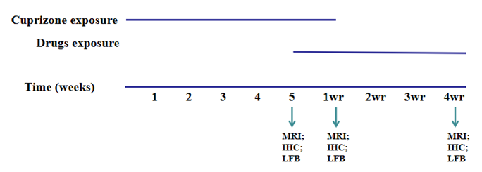 Figure. 1. Example of the cuprizone induced mouse model study paradigm
Our Capabilities
- We utilize cuprizone induced mouse model to explore effective therapeutic options for demyelination of the central nervous system.
- We detect CC demyelination by Luxol Fast Blue (LFB) staining and MRI.
- We quantitatively assess myelination of the corpus callosum through demyelination score system.
- We evaluate the number of oligodendrocytes, astrocytes, macrophages and microglia via IHC.
Assays Available
- Histopathological evaluation
- LFB staining
- MRI
With extensive experience in the field of MS, we are confident to help you to overcome any upcoming challenges. Our experts are fully capable of customizing our protocols and assays to meet your specific needs. With our help, we wish to facilitate your research with high efficiency.
Study Examples
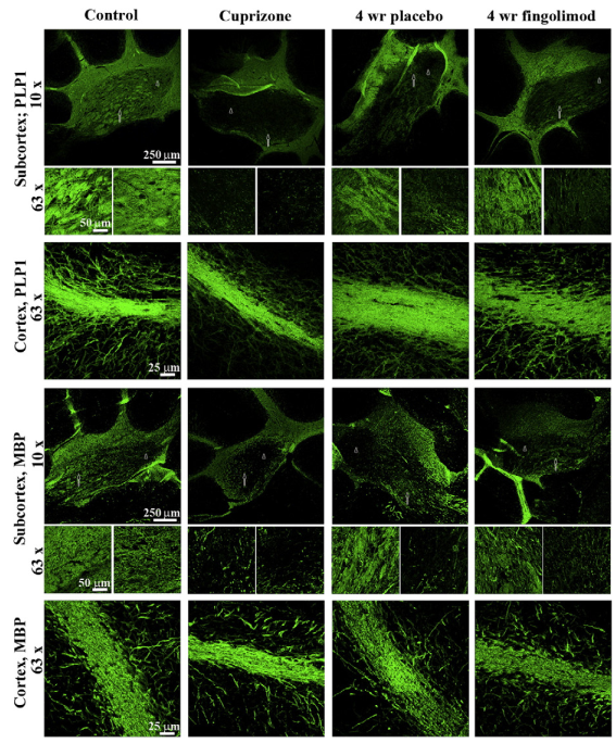 Figure. 2. Representative pictures show myelination (PLP1 and MBP) in the subcortex and cortex of mouse cerebellum for control, 5 weeks of cuprizone exposure and after 4 weeks of fingolimod/placebo treatment.
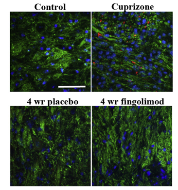 Figure. 3. Subcortical β-APP (red) and neurofilament (green) staining of control, after 5 weeks of cuprizone exposure and after 4 weeks of recovery.
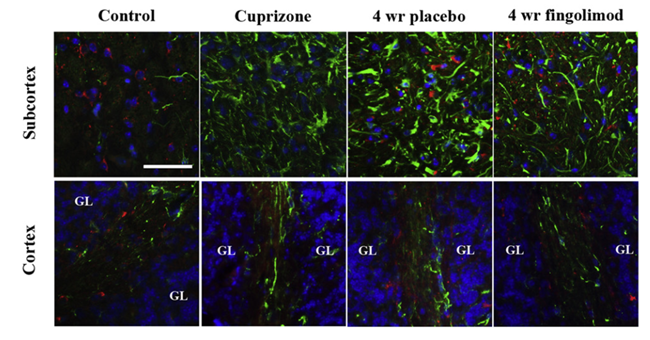 Figure. 4. NOGO-A positive mature oligodendrocytes (red) and GFAP-expressing astrocytes (green) in subcortex and cortex in control, 5 weeks of cuprizone exposure and after 4 weeks of recovery.
Quotation and ordering
If you have any special needs or questions regarding our services, please feel free to contact us. We look forward to cooperating with you in the future.
Reference
Maria Nordheim Alme, et al. Fingolimod does not enhance cerebellar remyelination in the cuprizone model[J]. Journal of Neuroimmunology, 2015, 285:180-186.
For research use only. Not for any other purpose.
Disease Models
- Oncology Models
-
Inflammation & Autoimmune Disease Models
- Rheumatoid Arthritis Models
- Glomerulonephritis Models
- Multiple Sclerosis (MS) Models
- Ocular Inflammation Models
- Sjögren's Syndrome Model
- LPS-induced Acute Lung Injury Model
- Peritonitis Models
- Passive Cutaneous Anaphylaxis Model
- Delayed-Type Hypersensitivity (DTH) Models
- Inflammatory Bowel Disease Models
- Systemic Lupus Erythematosus Animal Models
- Oral Mucositis Model
- Asthma Model
- Sepsis Model
- Psoriasis Model
- Atopic Dermatitis (AD) Model
- Scleroderma Model
- Gouty Arthritis Model
- Carrageenan-Induced Air Pouch Synovitis Model
- Carrageenan-Induced Paw Edema Model
- Experimental Autoimmune Myasthenia Gravis (EAMG) Model
- Graft-versus-host Disease (GvHD) Models
-
Cardiovascular Disease Models
- Surgical Models
- Animal Models of Hypertension
- Venous Thrombosis Model
- Atherosclerosis model
- Cardiac Arrhythmia Model
- Hyperlipoidemia Model
- Doxorubicin-induced Heart Failure Model
- Isoproterenol-induced Heart Failure Model
- Arterial Thrombosis Model
- Pulmonary Arterial Hypertension (PAH) Models
- Heart Failure with Preserved Ejection Fraction (HFpEF) Model
- Cardio-Renal-Metabolic (CKM) Syndrome Model
-
Neurological Disease Models
- Alzheimer's Disease Modeling and Assays
- Seizure Models
- Parkinson's Disease Models
- Ischemic Stroke Models
- Acute Spinal Cord Injury (ASCI) Model
- Traumatic Brain Injury (TBI) Model
- Hypoxic-Ischemic Encephalopathy (HIE) Model
- Tourette Syndrome (TS) Model
- Amyotrophic Lateral Sclerosis (ALS) Model
- Huntington's Disease (HD) Model
- Intracerebral hemorrhage (ICH) Models
- Schizophrenia Model
- Depression Models
- Pain Models
-
Metabolic Disease Models
- Type 1 Diabetes Mellitus Model
- Type 2 Diabetes Mellitus Model
- Animal Model of Hyperuricemia
-
Nonalcoholic Fatty Liver Disease Model
- High-Fat Diet-Induced Nonalcoholic Fatty Liver Disease (NAFLD) Model
- Methionine and Choline Deficient (MCD) Diet-Induced Nonalcoholic Fatty Liver Disease (NAFLD) Model
- Gubra-Amylin NASH (GAN) Diet-Induced Nonalcoholic Fatty Liver Disease (NAFLD) Model
- Streptozotocin (STZ) Induced Nonalcoholic Fatty Liver Disease (NAFLD) Model
- High Fat Diet-Induced Obesity Model
- Diabetic Foot Ulcer (DFU) Model
- Cardio-Renal-Metabolic (CKM) Syndrome Model
- Liver Disease Models
- Rare Disease Models
- Respiratory Disease Models
- Digestive Disease Models
-
Urology Disease Models
- Cisplatin-induced Nephrotoxicity Model
- Unilateral Ureteral Obstruction Model
- 5/6 Nephrectomy Model
- Renal Ischemia-Reperfusion Injury (RIRI) Model
- Diabetic Nephropathy (DN) Models
- Passive Heymann Nephritis (PHN) Model
- Adenine-Induced Chronic Kidney Disease (CKD) Model
- Kidney Stone Model
- Doxorubicin-Induced Nephropathy Model
- Orthotopic Kidney Transplantation Model
- Benign Prostatic Hyperplasia (BPH) Model
- Peritoneal Fibrosis Model
- Cardio-Renal-Metabolic (CKM) Syndrome Model
- Orthopedic Disease Models
- Ocular Disease Models
- Infectious Disease Models
- Skin Disease Models
- Otology Disease Models