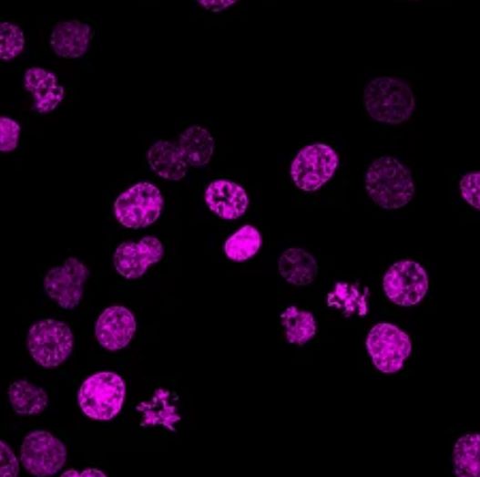
Fluorescent Nuclear Staining Dyes
Fluorescent nuclear staining dyes have revolutionized the field of cellular biology by providing researchers with powerful tools to visualize and study the intricate architecture of the cell nucleus. These dyes, such as DAPI (4',6-diamidino-2-phenylindole), Hoechst dyes, and SYTOX dyes, emit fluorescence upon binding to DNA or RNA, offering a profound insight into nuclear morphology, chromatin structure, and cellular dynamics.
Mechanisms of Nuclear Staining
The specificity of fluorescent nuclear staining dyes lies in their ability to selectively bind to nucleic acids within the cell nucleus. Upon permeating the cell membrane, these dyes interact with the phosphate backbone of DNA or RNA, leading to a distinctive emission of fluorescence upon excitation. The fluorescent signals generated by these dyes provide vital information about nuclear organization, DNA packaging, and cellular processes such as DNA replication and transcription. The mechanistic insight gained from nuclear staining techniques serves as a cornerstone for elucidating fundamental biological phenomena.
Features and Benefits of Fluorescent Nuclear Staining Dyes
Fluorescent nuclear staining dyes exhibit several advantageous features that render them indispensable for life science research. These include high affinity and specificity for nucleic acids, minimal cellular toxicity, stability in fixed and live cells, and compatibility with a range of imaging modalities. Additionally, the availability of a variety of fluorophores allows for multiplexing, enabling the simultaneous visualization of different cellular components and processes within the same sample.
- High affinity and specificity. These dyes exhibit high affinity and specificity for binding to nucleic acids within the cell nucleus. This targeted interaction allows for accurate labeling of nuclear DNA or RNA without non-specific interactions, ensuring precise visualization and analysis of nuclear structures.
- Minimal cellular toxicity. Many fluorescent nuclear staining dyes are designed to minimize cellular toxicity, enabling their use in live cell imaging and long-term studies without adversely affecting cellular viability or function.
- Stability and compatibility. These dyes demonstrate stability in fixed and live cells, making them suitable for a wide range of experimental conditions. Additionally, they are compatible with various imaging modalities, including fluorescence microscopy, confocal microscopy, and high-content screening platforms, allowing for versatile and multi-dimensional analysis of nuclear structures and dynamics.
- Multiplexing capabilities. The availability of a variety of fluorophores allows for multiplexing, which enables the simultaneous visualization of various cellular components within the same sample. This feature is particularly valuable in studying complex cellular processes involving multiple molecular targets or organelles.
- Reliability and reproducibility. Rigorous characterization and validation of these dyes ensure their reliability and reproducibility in experimental applications. This reliability is critical for generating consistent and meaningful data across different experiments and research studies.
Applications and Uses of Fluorescent Nuclear Staining Dyes
Fluorescent nuclear staining dyes find wide-ranging utility in cell biology research, serving as essential tools for a multitude of applications. These dyes are fundamental in visualizing nuclear morphology, including the delineation of nuclear size, shape, and substructures.
- Visualizing nuclear morphology. Fluorescent nuclear staining dyes are essential for visualizing nuclear morphology, including the size, shape, and substructures of the cell nucleus. This capability is foundational for studying nuclear organization and identifying changes in nuclear architecture associated with cellular processes or pathological conditions.
- Cell cycle analysis. These dyes facilitate precise cell cycle analysis by enabling the determination of DNA content in individual cells. This allows researchers to identify cells in different stages of the cell cycle, monitor cell cycle progression, and investigate factors influencing cell cycle regulation.
- Studying apoptosis and mitosis. Fluorescent nuclear staining dyes play a critical role in studying apoptosis and mitosis by enabling the visualization of nuclear condensation, fragmentation, and spindle formation. This capability is invaluable for elucidating the mechanisms and dynamics of these fundamental cellular processes.
- Assessing chromatin organization. These dyes are instrumental in assessing chromatin organization within the nucleus, providing insights into the spatial arrangements of DNA and histones. This is pivotal for understanding gene expression patterns, epigenetic modifications, and the regulation of chromatin structure.
- Live cell imaging and tracking cellular dynamics. In addition to fixed cell imaging, these dyes are employed in live cell imaging, permitting the real-time visualization of nuclear dynamics and cellular processes. This capability is essential for tracking cellular dynamics, cell migration, and studying dynamic interactions within the nucleus.
Creative Bioarray Relevant Recommendations
Creative Bioarray provides many fluorescent dyes that are highly specific to a variety of organelles and can be used to monitor cell health, cell death, metabolic activity, autophagy, cell tracking, cell migration, and invasion. Our team is always committed to providing customers with high-quality products and services.
| Cat. No. | Product Name |
| FDIR-D0094 | Hoechst 33258 |
| FDIR-D0095 | Hoechst 33342 |
| FDIR-D0096 | VitalTracker® Green Live Cell Nuclear Dye |
| FDIR-D0097 | VitalTracker® Red Live Cell Nuclear Dye |
| FDIR-D0098 | VitalTracker® FIR694 Live Cell Nuclear Dye |
| FDIR-D0103 | VitalTracker® FIR694 Fixed Cell Nuclear Dye |