Negative Staining TEM
Negative-stain transmission electron microscopy (TEM) is a technique that has provided nanometer resolution images of macromolecules for about 60 years. Isolated organelles, bacteria and viruses pose a problem when imaging in a TEM since they do not have high enough contrast (they are not electron-dense). For this purpose, Creative Bioarray uses negative staining. It is the perfect tool when it comes to size distribution of liposomes, exosomes or polymer nanoparticles.
Gene therapy offers great potential in the treatment and management of a plethora of diseases. Viral vectors (AAV, Adeno-virus, lentiviruses, etc.) are widely used delivery vectors in research and gene therapy. Negative stain TEM (nsTEM) provides detailed information about overall morphology of the viral vector samples and characterizes the properties such as the stability and purity. nsTEM analysis from Creative Bioarray provides comparable data when there are changes in the production conditions of upstream and downstream processes, or modifications to product formulations.
The following parameters of the viral vectors can be monitored using nsTEM:
- General morphology
- Integrity and debris/impurities
- Level of clusters/aggregation
- Size distribution
| Creative Bioarray We've Characterized | |
| Liposomes | Exosomes |
| Viruses, attenuated and recombinant | Extracellular Vesicles |
Virus-Like Particles (VLPs)
| |
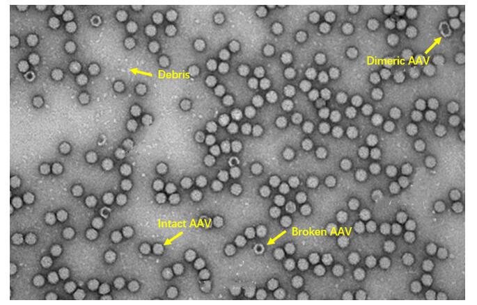
 Figure 1. nsTEM image of AAV particles. Internally stained AAV particles are broken or incorrectly assembled particles. The intact AAV particles appear as bright particles exhibiting a well-defined exterior. In the image it is also possible to identify the debris, as well as dimeric AAV particles.
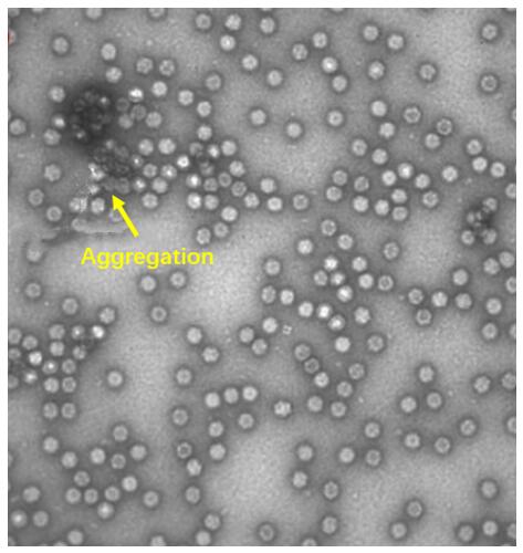
 Figure 2. nsTEM image of arregation AAV particles.
Features of Creative Bioarray's Negative Staining Service:
- Accurate - When the analysis is finished, we provide you with the TEM images and a detailed report
- Value - We focus on the quality of our service and all supported by competitive pricing
- Efficiency - We are able to provide the fastest turnaround time of any supplier in the industry
Quotation and ordering
Our customer service representatives are available 24hr a day! We thank you for choosing Creative Bioarray at your preferred Negative Staining TEM Services.
Explore Other Options
For research use only. Not for any other purpose.
Services
-
Cell Services
- Cell Line Authentication
- Cell Surface Marker Validation Service
-
Cell Line Testing and Assays
- Drug-Resistant Cell Models
- Cell Viability Assays
- Cell Proliferation Assays
- Cell Migration Assays
- Soft Agar Colony Formation Assay Service
- SRB Assay
- Cell Apoptosis Assays
- Cell Cycle Assays
- Cell Angiogenesis Assays
- DNA/RNA Extraction
- Cellular Phosphorylation Assays
- Stability Testing
- Sterility Testing
- Endotoxin Detection and Removal
- Phagocytosis Assays
- Cell-Based Screening and Profiling Services
- 3D-Based Services
- Custom Cell Services
- Cell-based LNP Evaluation
-
Stem Cell Research
- iPSC Generation
- iPSC Characterization
-
iPSC Differentiation
- Neural Stem Cells Differentiation Service from iPSC
- Astrocyte Differentiation Service from iPSC
- Retinal Pigment Epithelium (RPE) Differentiation Service from iPSC
- Cardiomyocyte Differentiation Service from iPSC
- T Cell, NK Cell Differentiation Service from iPSC
- Hepatocyte Differentiation Service from iPSC
- Beta Cell Differentiation Service from iPSC
- Brain Organoid Differentiation Service from iPSC
- Cardiac Organoid Differentiation Service from iPSC
- Kidney Organoid Differentiation Service from iPSC
- GABAnergic Neuron Differentiation Service from iPSC
- Undifferentiated iPSC Detection
- iPSC Gene Editing
- iPSC Expanding Service
- MSC Services
- Stem Cell Assay Development and Screening
- Cell Immortalization
-
Molecular Biology Solutions
- Chromosome & Genomic Analysis
-
Cytogenetics & Molecular Cytogenetics Analysis
- Fluorescent In Situ Hybridization (FISH)
- In Situ Hybridization (ISH) & RNAscope
- ImmunoFISH (FISH+IHC)
- I-FISH
- Splice Variant Analysis (FISH)
- RNA FISH in Plant
- mtRNA Analysis (FISH)
- Digital ISH Image Quantification and Statistical Analysis
- Telomere Length Analysis (Q-FISH)
- Telomere Length Analysis (qPCR assay)
- Droplet Digital PCR (ddPCR)
- QuantiGene Plex Assay
- Probe Development & Quality Control
- ISH/FISH Analysis for Therapeutic R&D
- Cell Line Characterization
- Pathogen & Microbial Analysis
- Histology Services
- Exosome Research Services
- Drug Metabolism and Pharmacokinetics (DMPK)
-
Safety Evaluation Services
- High-Throughput Toxicity Screening
- High-Content Cytotoxicity Screening
- In Vitro Cardiotoxicity
- In Vitro Genotoxicity
- Hepatotoxicity
- In Vitro Neurotoxicity
- In Vitro Nephrotoxicity
- In Vitro Dermal Toxicology
- Ocular Toxicity
- In Vitro Cytotoxicity
- Endocrine Disruption Screening Assay
- In Vivo Toxicity Study