Radiation-Induced Dermatitis Model
Radiation-induced dermatitis is a common and distressing side effect of radiation therapy, affecting nearly 90% of patients. This condition manifests as moderate-to-severe skin reactions, significantly diminishing the quality of life and potentially complicating the treatment process. Radiation not only damages the skin surface but also affects deeper tissues, leading to symptoms such as dryness, loss of elasticity, pigmentation changes, soft tissue fibrosis, capillary dilatation, and dermatitis in the treated areas. Furthermore, it causes irreversible damage to the microvascular network and endothelial cells of small blood vessels in the skin, resulting in prolonged healing times and increased susceptibility to infections.
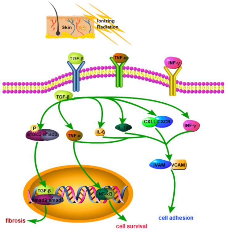 Fig. 1. Schematic diagram of related molecular mechanisms that may be involved in radiation-induced skin injury (RSI). (Yang et al. 2020)
To address these challenges, Creative Bioarray has developed the Radiation-Induced Dermatitis Model. This innovative model is designed to simulate the complex skin reactions triggered by radiation therapy, providing a critical tool for evaluating the efficacy of new therapeutic compounds aimed at alleviating these common side effects. By leveraging this model, researchers can gain deeper insights into the mechanisms of radiation-induced skin damage and accelerate the development of effective treatments to improve the quality of life for cancer patients undergoing radiation therapy.
Our Radiation-Induced Dermatitis Model
- Animal Species
Mouse
- Modeling Method
Mice are treated with an X-ray radiation dose of 40Gy to induce dermatitis.
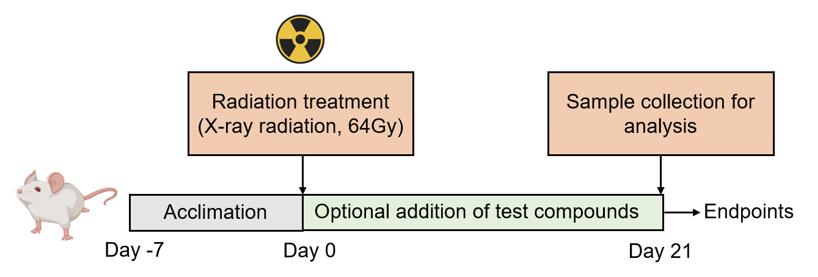 Fig. 2. Schematic diagram of the modeling method of Radiation-Induced Dermatitis Model.
- Endpoints
- In-life dermatitis evaluation
- Histology analysis: H&E staining, Masson staining
- Body weight
- qPCR or Western blot
- Other customized endpoints
Example Data
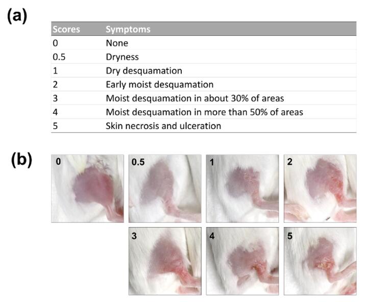 Fig. 3. Radiation dermatitis scores in a BALB/c mouse model: (a) Definitions of the radiation dermatitis scores and (b) photographs of a mouse hind leg representing the radiation dermatitis scores. (Yang et al. 2020)
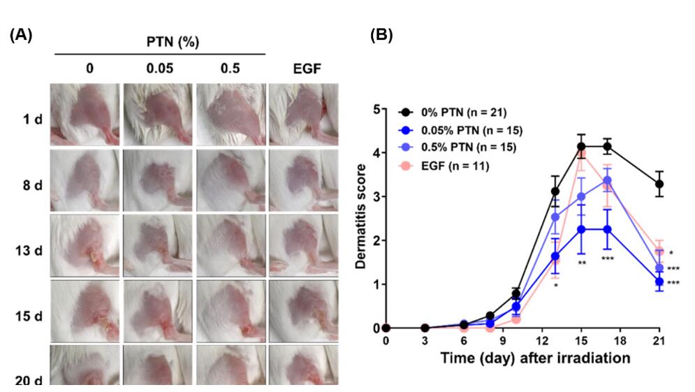 Fig. 4. Effects of the topical application of phlorotannins (PTNs) on radiation dermatitis: (a) Representative photographs of the irradiated skin treated with topical PTNs or rhEGF. (b) Time-course of changes in RD score. (Yang et al. 2020)
Quotation and Ordering
By partnering with Creative Bioarray, you gain access to a robust solution that not only accelerates your drug discovery process but also ensures the rigorous evaluation and optimization of your compounds for superior efficacy and safety. If you are interested in our services, please feel free to contact us at any time or submit an inquiry to us directly.
References
- Yang, K., et al. Topical application of phlorotannins from brown seaweed mitigates radiation dermatitis in a mouse model. Marine drugs, 2020, 18(8): 377.
- Yang, X., et al. Radiation-induced skin injury: pathogenesis, treatment, and management[J]. Aging (Albany NY), 2020, 12(22): 23379.
For research use only. Not for any other purpose.
Disease Models
- Oncology Models
-
Inflammation & Autoimmune Disease Models
- Rheumatoid Arthritis Models
- Glomerulonephritis Models
- Multiple Sclerosis (MS) Models
- Ocular Inflammation Models
- Sjögren's Syndrome Model
- LPS-induced Acute Lung Injury Model
- Peritonitis Models
- Passive Cutaneous Anaphylaxis Model
- Delayed-Type Hypersensitivity (DTH) Models
- Inflammatory Bowel Disease Models
- Systemic Lupus Erythematosus Animal Models
- Oral Mucositis Model
- Asthma Model
- Sepsis Model
- Psoriasis Model
- Atopic Dermatitis (AD) Model
- Scleroderma Model
- Gouty Arthritis Model
- Carrageenan-Induced Air Pouch Synovitis Model
- Carrageenan-Induced Paw Edema Model
- Experimental Autoimmune Myasthenia Gravis (EAMG) Model
- Graft-versus-host Disease (GvHD) Models
-
Cardiovascular Disease Models
- Surgical Models
- Animal Models of Hypertension
- Venous Thrombosis Model
- Atherosclerosis model
- Cardiac Arrhythmia Model
- Hyperlipoidemia Model
- Doxorubicin-induced Heart Failure Model
- Isoproterenol-induced Heart Failure Model
- Arterial Thrombosis Model
- Pulmonary Arterial Hypertension (PAH) Models
- Heart Failure with Preserved Ejection Fraction (HFpEF) Model
- Cardio-Renal-Metabolic (CKM) Syndrome Model
-
Neurological Disease Models
- Alzheimer's Disease Modeling and Assays
- Ischemic Stroke Models
- Acute Spinal Cord Injury (ASCI) Model
- Traumatic Brain Injury (TBI) Model
- Hypoxic-Ischemic Encephalopathy (HIE) Model
- Tourette Syndrome (TS) Model
- Amyotrophic Lateral Sclerosis (ALS) Model
- Huntington's Disease (HD) Model
- Intracerebral hemorrhage (ICH) Models
- Schizophrenia Model
- Depression Models
- Pain Models
-
Metabolic Disease Models
- Type 1 Diabetes Mellitus Model
- Type 2 Diabetes Mellitus Model
- Animal Model of Hyperuricemia
-
Nonalcoholic Fatty Liver Disease Model
- High-Fat Diet-Induced Nonalcoholic Fatty Liver Disease (NAFLD) Model
- Methionine and Choline Deficient (MCD) Diet-Induced Nonalcoholic Fatty Liver Disease (NAFLD) Model
- Gubra-Amylin NASH (GAN) Diet-Induced Nonalcoholic Fatty Liver Disease (NAFLD) Model
- Streptozotocin (STZ) Induced Nonalcoholic Fatty Liver Disease (NAFLD) Model
- High Fat Diet-Induced Obesity Model
- Diabetic Foot Ulcer (DFU) Model
- Liver Disease Models
- Rare Disease Models
- Respiratory Disease Models
- Digestive Disease Models
-
Urology Disease Models
- Cisplatin-induced Nephrotoxicity Model
- Unilateral Ureteral Obstruction Model
- 5/6 Nephrectomy Model
- Renal Ischemia-Reperfusion Injury (RIRI) Model
- Diabetic Nephropathy (DN) Models
- Passive Heymann Nephritis (PHN) Model
- Adenine-Induced Chronic Kidney Disease (CKD) Model
- Kidney Stone Model
- Doxorubicin-Induced Nephropathy Model
- Orthotopic Kidney Transplantation Model
- Benign Prostatic Hyperplasia (BPH) Model
- Peritoneal Fibrosis Model
- Orthopedic Disease Models
- Ocular Disease Models
- Infectious Disease Models
- Skin Disease Models
- Otology Disease Models