
Skeletal Cells
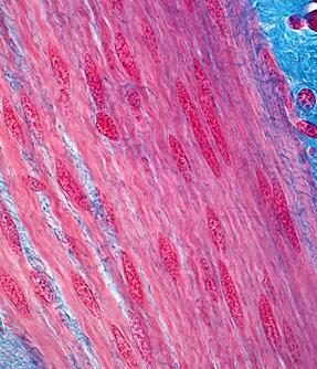

The musculoskeletal system is composed of interconnected tissues that provide structural support, movement, mechanical stability, and tissue repair throughout the body. Coordinated interactions among skeletal muscle cells, chondrocytes, synovial cells, tendon cells, and intervertebral disc cells are essential for maintaining normal musculoskeletal function and responding to injury or degeneration.
Our skeletal cell portfolio includes primary human muscle, cartilage, synovial, tendon, and intervertebral disc cell models, together with disease-specific and fluorescent reporter cell variants. These cell resources support research in musculoskeletal biology, osteoarthritis, rheumatoid arthritis, muscular dystrophy, regenerative medicine, tissue engineering, and therapeutic development.

5
Musculoskeletal Tissue
Categories

100 %
Mycoplasma-Free
Tested

4 +
Disease Models
Available
Filter By:
Our Skeletal Cell Portfolio Highlights

Comprehensive Musculoskeletal Cell Resources
Our collection represents key cellular components involved in muscle function, joint biology, cartilage maintenance, and connective tissue remodeling.
-
Skeletal Muscle Cell Models
- Skeletal muscle cells and myoblasts
- Satellite cells and muscle progenitor cells
- Microvascular endothelial cells associated with skeletal muscle
-
Joint and Cartilage Cell Models
- Articular chondrocytes
- Normal, osteoarthritis, and rheumatoid arthritis synoviocytes
- Reporter-labeled chondrocyte models
-
Connective Tissue and Disc Models
- Tenocytes
- Nucleus pulposus cells
- Annulus fibrosus cells

Applications in Musculoskeletal Research
These models enable studies of tissue development, degeneration, regeneration, disease progression, and therapeutic intervention.
-
Muscle Biology & Regeneration
Investigate muscle development, satellite cell activation, tissue repair, and mechanisms regulating skeletal muscle regeneration.
-
Joint and Cartilage Disease Research
Support studies of osteoarthritis, rheumatoid arthritis, cartilage degeneration, and inflammatory joint disorders.
-
Muscular Dystrophy & Metabolic Disease Models
Utilize DMD and diabetes-associated muscle cell models to investigate disease mechanisms and therapeutic strategies.
-
Tissue Engineering & Regenerative Medicine
Facilitate development of engineered musculoskeletal tissues, biomaterials, and regenerative therapies.
What are skeletal muscle satellite cells commonly used for?
Satellite cells are resident muscle stem cells responsible for muscle growth and repair. They are widely used to study muscle regeneration, stem cell biology, and muscle disease mechanisms.
How do synoviocytes support arthritis research?
Synoviocytes regulate joint homeostasis and inflammatory responses within the synovial membrane. Disease-specific synoviocyte models are valuable for studying osteoarthritis and rheumatoid arthritis pathogenesis.
What applications are chondrocytes used for?
Chondrocytes are the primary cells responsible for maintaining cartilage structure and extracellular matrix production, making them essential for cartilage biology and degeneration studies.
What are nucleus pulposus and annulus fibrosus cells used for?
These intervertebral disc cell types are commonly used to investigate disc degeneration, extracellular matrix remodeling, spinal disorders, and regenerative approaches for disc repair.
How can DMD muscle cell models support translational research?
Duchenne muscular dystrophy-derived muscle cells provide clinically relevant platforms for studying disease mechanisms, biomarker discovery, gene therapies, and novel therapeutic interventions.
What advantages do fluorescently labeled chondrocytes provide?
GFP- and RFP-expressing chondrocytes facilitate live-cell imaging, cell tracking, migration analysis, and visualization of cartilage-related cellular interactions in vitro.