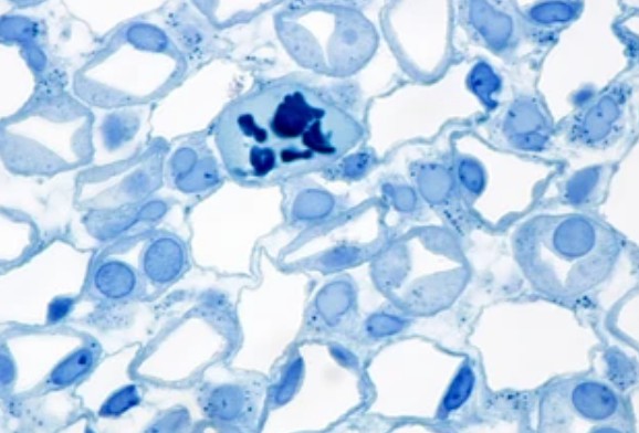
Comparison of Membrane Stains vs. Cell Surface Stains
In biological research, the visualization and characterization of cell membranes and cell surface markers play a crucial role in understanding cellular structure, function, and interactions. Membrane stains and cell surface stains are valuable tools that enable researchers to study the composition, dynamics, and behavior of cell membranes and surface proteins.

Membrane Stains
Membrane stains are substances that selectively bind to cell membranes, allowing researchers to observe and analyze the structure and properties of the cell membrane. There are various types of membrane stains available, including lipophilic dyes and fluorescent probes.
- Lipophilic dyes. They are a class of membrane stains that possess an affinity for lipid-rich environments, such as cell membranes. These dyes can be incorporated into the lipid bilayer of the cell membrane, resulting in fluorescent staining. One commonly used lipophilic dye is DiI (1,1'-dioctadecyl-3,3,3',3'-tetramethylindocarbocyanine perchlorate), which emits a red fluorescence upon binding to the cell membrane. Lipophilic dyes are known for their long-lasting staining and are particularly useful for tracking cell migration and cell-cell interactions.
- Fluorescent probes. They are another type of membrane stain that can selectively bind to cell membranes. These probes are often small molecules conjugated with fluorophores, which emit fluorescence upon binding to the cell membrane. Examples of fluorescent probes include FM dyes, which are used for studying endocytosis and vesicle recycling, and membrane-permeant DNA dyes, such as Hoechst stains, which enable visualization of DNA within the cell nucleus. Fluorescent probes provide researchers with the ability to study membrane dynamics and cellular processes in real time.
Cell Surface Stains
Cell surface stains are specifically designed to target and label cell surface markers, such as proteins or carbohydrates, allowing for the identification and characterization of specific cell populations. They play a crucial role in biological research, particularly in studying cell surface markers, cellular functions, and interactions.
- Fluorescent dyes. They are widely used to label and visualize cell surface proteins. Examples include fluorescein isothiocyanate (FITC), rhodamine, Texas Red, Alexa Fluor dyes, and cyanine dyes (Cy3, Cy5, etc.). These dyes can be conjugated to antibodies or other molecules that specifically bind to cell surface proteins.
- Antibodies. They can specifically target and stain cell surface proteins, which can be conjugated to fluorescent dyes or enzymes for visualization. Examples include primary antibodies that recognize specific cell surface receptors or adhesion molecules, such as CD markers.
Comparative Analysis of Membrane Stains vs. Cell Surface Stains
| Comparative items | Membrane stains vs. cell surface stains | |
| Specificity and selectivity | Membrane stains, such as lipophilic dyes and fluorescent probes, are designed to selectively label the cellular membranes based on their lipid composition. These stains are highly specific and enable the visualization of membrane structures and dynamics. | In contrast, cell surface stains, including immunofluorescence and lectin staining, target specific antigens or glycoproteins present on the outer surface of the cell membrane. These stains provide exquisite specificity, allowing for the identification and characterization of surface markers and cellular interactions. |
| Visualization and imaging | Membrane stains are primarily employed for visualizing the overall structure and organization of cellular membranes. They are instrumental in studying membrane dynamics, lipid rafts, and intracellular trafficking. | Cell surface stains are specifically tailored to visualize and study the expression patterns, localization, and distribution of surface proteins and carbohydrates, providing insights into cellular functions and interactions within complex biological systems. |
| Techniques and methodologies | Techniques such as lipid bilayer labeling and membrane potential dyes are utilized to study cellular membrane properties, including integrity, fluidity, and permeability. | Techniques like immunofluorescence, flow cytometry, and lectin staining are commonly employed to analyze cell surface markers, receptor expression, and antigen presentation, offering valuable information on cell identity and behavior. |
Applications of Membrane and Cell Surface Stains in Medical Research
- Cancer research. Membrane and cell surface stains are valuable tools in cancer research for identifying and characterizing cancer cells. By targeting specific cell surface markers associated with cancer, researchers can distinguish cancer cells from normal cells, analyze tumor heterogeneity, and study cancer progression.
- Neuroscience. Membrane stains and cell surface stains are essential in neuroscience research for studying neuronal morphology, synapse formation, and cell-cell communication. Lipophilic dyes, for example, are used to trace neuronal pathways and study neuronal connectivity.
Creative Bioarray Relevant Recommendations
At Creative Bioarray, our comprehensive range of stains is meticulously designed to empower researchers with precise tools for visualizing and studying cellular membrane structures and dynamics.
| Cat. No. | Product Name |
| FDIR-D0109 | DiA |
| FDIR-D0111 | DiBAC4 |
| FDIR-D0112 | DiI |
| FDIR-D0113 | DiD |
| FDIR-D0114 | DiR |
| FDIR-D0115 | VitalTracker® Blue Cytoplasmic Membrane Dye |
| FDIR-D0116 | VitalTracker® Green Cytoplasmic Membrane Dye |
| FDIR-D0117 | VitalTracker® Orange Cytoplasmic Membrane Dye |
| FDIR-D0118 | VitalTracker® Red Cytoplasmic Membrane Dye |