
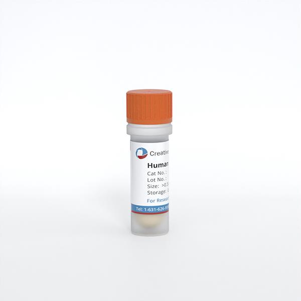
Human Keratinocytes - adult (HKs-a)
Cat.No.: CSC-C4005X
Species: Human
Source: Epidermis; Skin
Cell Type: Keratinocyte
- Specification
- Background
- Scientific Data
- Q & A
- Customer Review
Human Keratinocytes - adult (HKs-a) from Creative Bioarray are isolated from the Dermis of adult skin. The cells are cryopreserved at Passage 2 and delivered frozen. Each vial contains at least 0.5 *10^6 cells. HKs-a are negative for HIV-1, HBV, HCV, mycoplasma, bacteria, yeast and fungi. Human Keratinocytes - adult (HKs-a) are guaranteed to at 15 population doublings under the conditions provided by Creative Bioarray. Repeated freezing and thawing of cells is not recommended.
Human keratinocytes - adult (HKs-a) are primary epidermal keratinocytes isolated from normal adult human skin. They retain key keratinocyte markers, including p63, cytokeratin 5/14 in basal cells and cytokeratin 1/10, involucrin, filaggrin, and loricrin upon differentiation. Cultured in low-calcium, serum-free medium, HKs-a maintain a proliferative basal phenotype; calcium switch or air-liquid interface (ALI) culture induces terminal differentiation and stratified epidermal architecture. This enables functional studies of epidermal barrier formation, cornification, tight junctions, and lipid organization.
Compared with neonatal keratinocytes or immortalized lines, HKs-a better reflect adult skin physiology, including matured barrier properties, inflammatory signaling, and donor-specific genetic background. They are highly relevant for modeling wound healing, psoriasis, atopic dermatitis, contact dermatitis, photoaging, and skin carcinogenesis, and for evaluating drugs, cosmetics, irritants, sensitizers, and wound-care products. Their compatibility with monolayer, 3D organotypic skin equivalents, co-culture with fibroblasts/immune cells, and high-content imaging supports mechanism and toxicity studies.
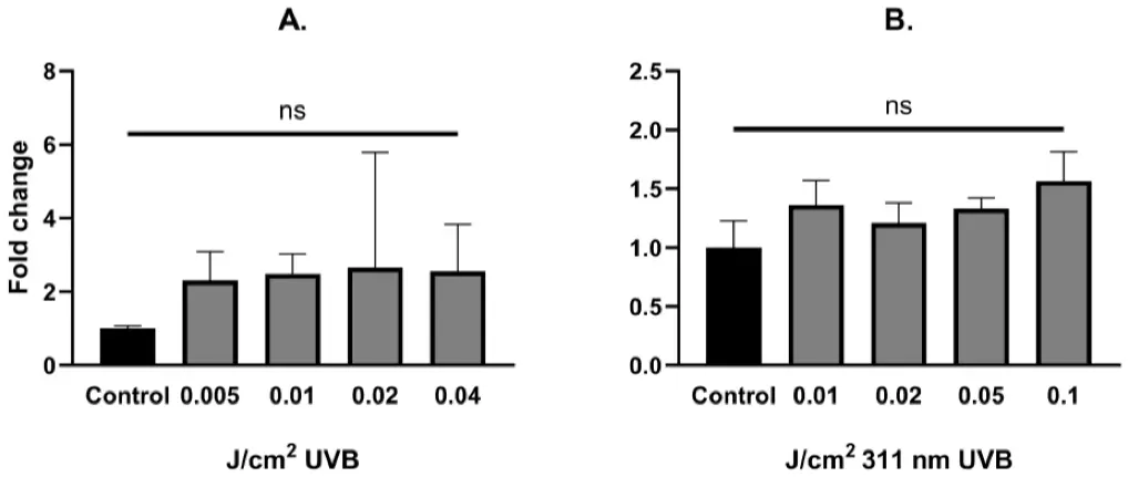
Ultraviolet B Exposure Does Not Influence the Expression of YAP mRNA in Human Epidermal Keratinocytes
The causal relationship between exposure to ultraviolet radiation and the development of skin cancers, as well as the role of YAP in cancer initiation and progression, implies that UVB could activate YAP or suppress Hippo pathway signaling. In order to address this issue, we focused on how different doses of UVB affect Hippo signaling pathway components and its upstream regulators, JNK1/2 and ABL1, in human keratinocytes. Moreover, we decided to determine how silencing YAP influences Hippo pathway component expression under the same study conditions.
UVB irradiation did not change the levels of YAP mRNA expression in human epidermal keratinocytes (Fig. 1A, B). LATS1, LATS2, ABL1 and MAP4K4 mRNA expression was significantly upregulated after UVB irradiation in non-YAP-silenced keratinocytes in a dose-dependent manner. After YAP silencing, only LATS2 and ABL1 showed significant mRNA upregulation. Regardless of YAP silencing, no changes in JNK1 and JNK2 mRNA expression were noted after irradiation. YAP mRNA expression does not significantly increase after exposure to UVB; however, it upregulates the expression of its proven (LATS1/2, JNK1/2) regulators, suggesting that in real-life settings, UV-induced dysregulation of the Hippo pathway may not be limited to YAP.
Ask a Question
Write your own review
Description: Keratinocytes are the primary type of cell found in the epidermis, the outermost layer of the skin. Keratinocytes represent the major cell type of the epidermis, making up about 90% of the cells. ...
Description: Diseased Human Epidermal Keratinocytes (D-HEK) are isolated from human donors that have been diagnosed with diabetes type II disease. Further donor information, including medications, BMI, and ...
Description: Keratinocytes are the primary type of cell found in the epidermis, the outermost layer of the skin. Keratinocytes represent the major cell type of the epidermis, making up about 90% of the cells. ...
Description: Bioarray epidermal keratinocytes are available from adult and neonatal donor tissue and are provided at the earliest passage without the use of a murine feeder layer. Keratinocytes are available from ...