Jurkat
Cat.No.: CSC-C9108W
Species: Homo sapiens (Human)
Source: Blood; Peripheral Blood
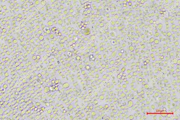
Morphology: round cells
- Specification
- Background
- Scientific Data
- Q & A
- Customer Review
- Documents
Jurkat cells are a well-known and extensively studied line of T lymphocyte (T-cell) cells, which were established from the peripheral blood of a 14-year-old male patient who was experiencing the first relapse of acute lymphoblastic leukemia (ALL) in 1976. This cell line is significant in immunological and cancer research, serving as a valuable model for studying T-cell leukemia and lymphoma.
Jurkat cells have been pivotal in various research areas, including immunology, virology, cancer biology, and drug development. They have been utilized in studies related to T-cell signaling, immune response modulation, viral infection mechanisms, and the development of novel therapies for leukemia and other T-cell malignancies. Their use has contributed to a deeper understanding of T-cell biology and the molecular mechanisms associated with leukemia and other related diseases.
Additionally, due to their well-characterized genetic background and established relevance to T-cell malignancies, Jurkat cells continue to be an essential tool in research aimed at the development of targeted therapies and the exploration of immunotherapeutic strategies.
Mitochondria Transfer Contributing to MSC-Induced Chemoresistance in T-All Cells
Despite the high cure rate of T cell acute lymphoblastic leukemia (T-ALL), drug resistance to chemotherapy remains a significant clinical problem. The study explored the potential mechanism by which bone marrow mesenchymal stem cells protect leukemia cells from the effects of chemotherapy.
In vitro, MSCs and Jurkat cells were cocultured. MSCs were labeled with a green fluorescent protein (GFP), and Jurkat cells were labeled with the mitochondria-specific dye MitoTracker Red. After 2 days of coculture, we quantified mitochondria transfer by flow cytometry. The results showed that 32.20 ± 5.21% (ara-C-treated group) or 30.00 ± 4.31% (MTX-treated group) of GFP-labeled MSCs were Red+, indicating that approximately 30% of the MSCs received mitochondria from Jurkat cells (Fig. 1a). However, only 0.59 ± 0.14% (ara-C-treated group) or 0.62 ± 0.15% (MTX-treated group) of the Jurkat cells were Red+ after 2 days of co-culture, indicating that few Jurkat cells received mitochondria from MSCs (Fig. 1b). These results showed that Jurkat cells could transfer mitochondria to MSCs when treated with chemotherapeutic drugs.
Whether mitochondria transfer could lead to drug resistance was examined in Jurkat cells. Annexin V/PI flow cytometry analysis and a cell viability assay showed that Jurkat cells treated with cytochalasin D had an increased apoptosis rate (Fig. 2c, d) and decreased cell viability (Fig. 2e), indicating that blocking mitochondria transfer decreased the capacity of MSCs to protect Jurkat cells from drug cytotoxicity. These results demonstrate that mitochondria transfer contributes to the MSC-induced chemoresistance of Jurkat cells. Taken together, the study found Jurkat cells transfer mitochondria to MSCs but receive few mitochondria from MSCs, resulting in chemoresistance.
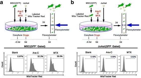 Fig. 1 Jurkat cells transfer mitochondria to MSCs when exposed to ara-C or MTX. (Wang J, et al., 2018)
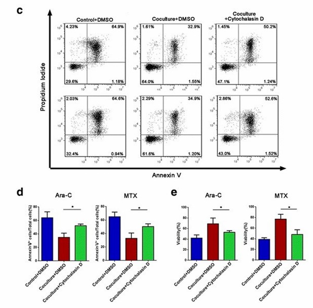 Fig. 2 Jurkat cells transfer mitochondria to MSCs when exposed to ara-C or MTX. (Wang J, et al., 2018)
Calcium Enhancing Transfection Efficiency in Jurkat Cells
Jurkat has been employed as an excellent surrogate model of human primary T-cells. However, presumably due to its T-cell origin, Jurkat cells are very difficult to transfect. It is reported that a simple addition of calcium ions (Ca2+) into culture media at optimal concentrations can enhance the efficiency of the polyplex-mediated transfection using poly (ethylene imine) (PEI).
Compared to the cell-only (CO) group, PEI polyplex without any calcium chloride (0 mM) induced GFP expression in a very small population of around 0.6% (Fig. 3a, e). A wide spectrum of GFP expression levels appears in 10- and 25-mM groups only (Fig. 3e), which was verified by the statistically significant increases in the mean fluorescence intensity (MFI) values of these groups (Fig. 3b). The GFP expression in the Jurkat cells were examined by confocal laser scanning microscope (CLSM) imaging at a high resolution (Fig. 3d). It is noteworthy that the numbers of live cells in 10 mM and 25 mM groups were significantly lower than 0 mM, presumably due to combined cytotoxicity of calcium and polyplex (Fig. 3c). Nevertheless, it is demonstrated that the addition of calcium chloride at concentrations of 10 mM or above can enhance the transfection efficiency of pDNA using PEI-based polyplexes on otherwise hard-to-transfect Jurkat T cells.
In direct comparisons, magnesium chloride groups did not show any effect in transfection compared to polyplex-only control (0 mM), while the addition of sodium chloride at indicated concentrations made the size of the GFP+ population rather decreased (Fig. 4a, b). Unlike the wide spectrum of GFP expression levels observed in the calcium chloride groups, most of the GFP + cells in the magnesium chloride groups resemble the low-level GFP expression of 0 mM control (Fig. 4a, c). Altogether, calcium cation instead of chloride anion seems to be responsible for the enhancement in polyplex transfection efficiency, and the other tested cations, Mg2+ and Na+, were ineffective.
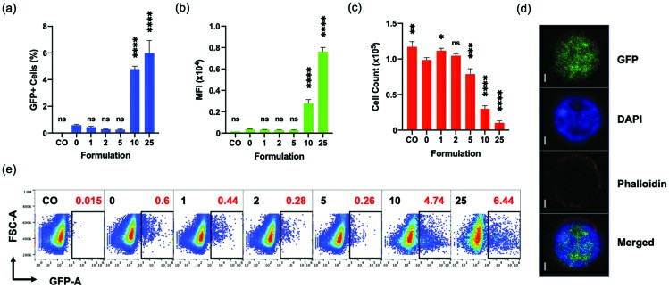 Fig. 3 Effect of CaCl2 on the transfection of Jurkat cells analyzed by flow cytometer at 96 hpt. (Ayyadevara VSSA and Roh KH, 2020)
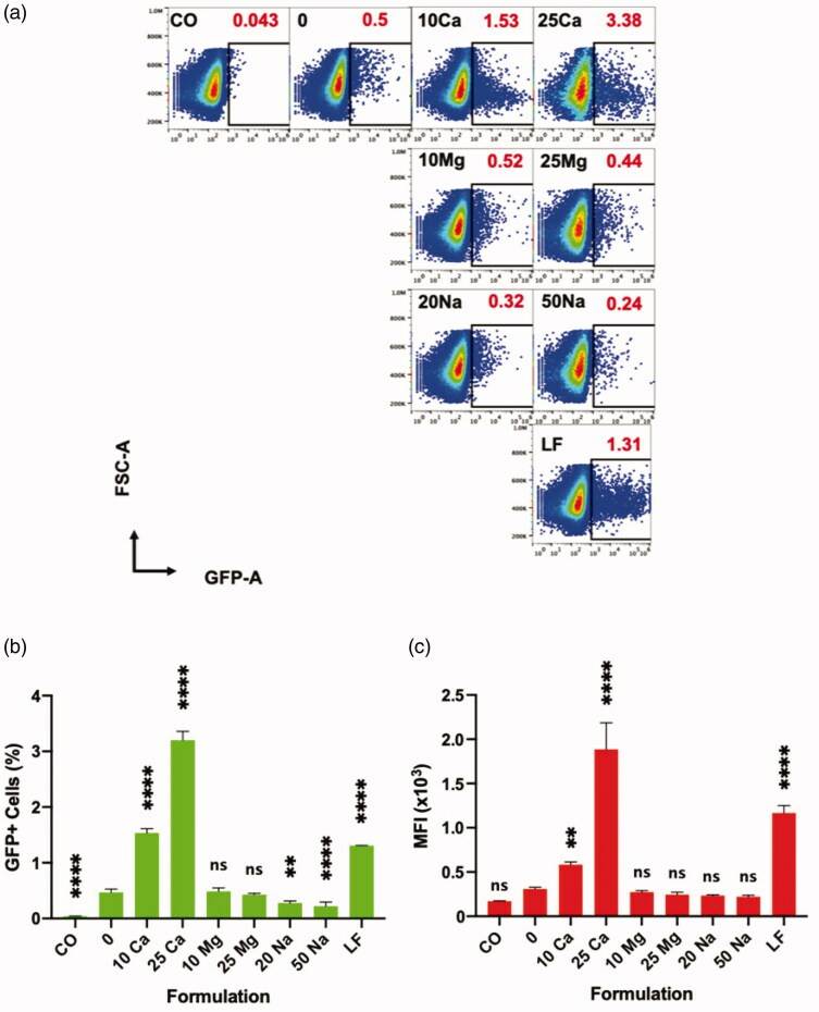 Fig. 4 Effect of various cations on the transfection of polyplex on Jurkat cells, and their direct comparison to lipoplex analyzed by flow cytometer at 96 h. (Ayyadevara VSSA and Roh KH, 2020)
Ask a Question
Write your own review
- You May Also Need

Description: Established in 2007 from the bone marrow mononuclear cells of an 82-year-old Japanese man with diffuse large B-cell lymphoma in the leukemic phase

Description: Established from the bone marrow of a 28-year-old man who developed the terminal leukemic phase of lymphosarcoma in 1976

Description: This cell line was derived from the bone marrow aspirate of a 59 year old male with erythroleukemia that became acute myelogenous leukaemia.The cells form colonies in soft-agar in the presence of ...

Description: Established from the pleural effusion of a 24-year-old woman with recurrent anaplastic large cell lymphoma (ALCL); cells were described to clonally derive from T-lineage lymphoid cells and to be ...

Description: Established from a 37-year-old man at second (refractory/terminal) relapse of Hodgkin lymphoma (nodular sclerosing -> lymphocyte depleted/stage IIISA -> stage IV) after both combined chemo- and ...

Description: Established from the peripheral blood of a 10-year-old Caucasian boy with acute lymphoblastic leukemia (pre B-ALL) at diagnosis in 1993
- Adipose Tissue-Derived Stem Cells
- Human Neurons
- Mouse Probe
- Whole Chromosome Painting Probes
- Hepatic Cells
- Renal Cells
- In Vitro ADME Kits
- Tissue Microarray
- Tissue Blocks
- Tissue Sections
- FFPE Cell Pellet
- Probe
- Centromere Probes
- Telomere Probes
- Satellite Enumeration Probes
- Subtelomere Specific Probes
- Bacterial Probes
- ISH/FISH Probes
- Exosome Isolation Kit
- Human Adult Stem Cells
- Mouse Stem Cells
- iPSCs
- Mouse Embryonic Stem Cells
- iPSC Differentiation Kits
- Mesenchymal Stem Cells
- Immortalized Human Cells
- Immortalized Murine Cells
- Cell Immortalization Kit
- Adipose Cells
- Cardiac Cells
- Dermal Cells
- Epidermal Cells
- Peripheral Blood Mononuclear Cells
- Umbilical Cord Cells
- Monkey Primary Cells
- Mouse Primary Cells
- Breast Tumor Cells
- Colorectal Tumor Cells
- Esophageal Tumor Cells
- Lung Tumor Cells
- Leukemia/Lymphoma/Myeloma Cells
- Ovarian Tumor Cells
- Pancreatic Tumor Cells
- Mouse Tumor Cells