THP-1 h
Cat.No.: CSC-6319W
Species: Homo sapiens (Human)
Source: Blood; Peripheral Blood
Morphology: continuous culture, grown in suspension, morphology large, round, single cells
Culture Properties: suspension
- Specification
- Background
- Scientific Data
- Q & A
- Customer Review
Tissue: peripheral blood;
Tumor: leukemia, acute monocytic;
Derived from: THP-1
vWA: 16;
FGA: 24,25;
TH01: 8,9.3;
D18S51: 13,14;
D21S11: 30,31.2;
D8S1179: 10,14;
Multiplex PCR: tested against human, rat, mouse primers.
Confirmed as human with cytochrome c oxidase subuni
The THP-1h cell line is a highly characterized and functionally enhanced human monocytic model, specifically selected for its superior responsiveness and phenotypic stability compared to standard monocytic lineages. Derived from a patient with acute monocytic leukemia, this "High-Response" (h) variant serves as a critical in vitro tool for researchers who require consistent, robust performance in inflammatory signaling, macrophage polarization, and vaccine development assays.
- Enhanced Differentiation Kinetics: THP-1h cells exhibit an accelerated and highly synchronized transition from monocytes to macrophage-like cells upon induction (e.g., with PMA). This leads to a more uniform cell population, providing higher sensitivity for studying M1/M2 macrophage polarization and tissue-specific immunity.
- Superior Inflammasome Sensitivity: Our THP-1h variant is validated for its robust activation of the NLRP3 inflammasome pathway. It demonstrates significantly higher secretion levels of pro-inflammatory cytokines like IL-1β and TNF-α following TLR-4 priming, making it the ideal substrate for screening small-molecule anti-inflammatory inhibitors.
- Optimal Phagocytic and Chemotactic Activity: These cells maintain high expression of essential surface markers such as CD14, CD11b, and CCR2. This ensures superior performance in functional assays, including pathogen phagocytosis, efferocytosis, and chemotactic migration studies.
- High Post-Thaw Recovery & Purity: Each lot is rigorously screened for exceptional post-thaw viability (>90%) and genetic stability. Their predictable doubling time and consistent morphology minimize experimental drift, ensuring that your high-throughput screening (HTS) data remains reproducible across multiple passages.
By utilizing the THP-1h cell line, you gain a high-fidelity, human-derived system that offers the sensitivity and reliability required for the most demanding preclinical research and drug discovery applications.
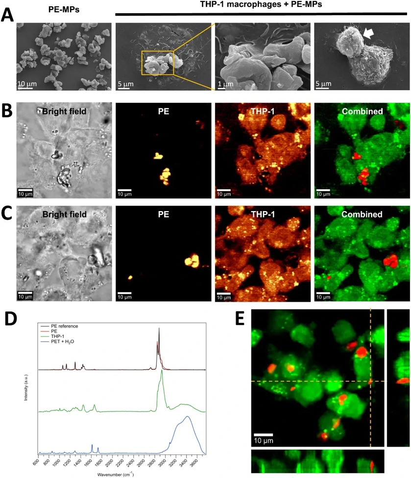
Polyethylene Microplastic Mixtures Activates THP-1 Macrophages with Inflammatory Features
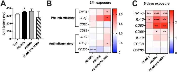
Microplastics (MPs) are pervasive in the environment and are toxic due to the nature of their polymers and associated additives, such as bisphenols (BPs) and per- and polyfluoroalkyl substances (PFAS). Nonetheless, there is a scarcity of in vitro studies investigating the toxicity of MPs combined with these additives and deciphering the underlying mechanisms in the context of recurrent exposures, which more accurately reflect human contamination. The objective of this study is to assess the differential toxic impacts of polyethylene microplastics (PE-MPs, 100 μg/mL) and a mixture of additives (i.e., AddMix containing 10 μM of BPA, BPS, PFOS, and PFOA) on naive THP-1 macrophages during unique or three repeated exposures over five days.
Scanning electron and Raman microscopy images indicated an effective internalization of PE-MPs by macrophages after 30 minutes and 24 hours of exposure. Three repeated exposures of THP-1 macrophages to PE-MPs and PE-MPs+AddMix over five days increased mitochondrial ROS production by 19 % and 13 %, respectively, and led to a more pronounced glycolytic phenotype, associated with the enhanced secretion of the pro-inflammatory cytokine IL-1β by 38 % with PE-MPs. Analysis of transcriptomic data revealed that PE-MPs and/or AddMix significantly enhanced the expression of genes primarily related to inflammation and modulation of lipid metabolism pathways. Our work highlights that MPs mixtures with additives, particularly upon repeated exposure, are recognized by macrophages as activation signals, leading to stimulation of inflammatory pathways, which require further investigation to be fully deciphered.
Ask a Question
Write your own review
- You May Also Need

Description: Established in 2007 from the bone marrow mononuclear cells of an 82-year-old Japanese man with diffuse large B-cell lymphoma in the leukemic phase

Description: Established from the bone marrow of a 28-year-old man who developed the terminal leukemic phase of lymphosarcoma in 1976

Description: This cell line was derived from the bone marrow aspirate of a 59 year old male with erythroleukemia that became acute myelogenous leukaemia.The cells form colonies in soft-agar in the presence of ...

Description: Established from the pleural effusion of a 24-year-old woman with recurrent anaplastic large cell lymphoma (ALCL); cells were described to clonally derive from T-lineage lymphoid cells and to be ...

Description: Established from a 37-year-old man at second (refractory/terminal) relapse of Hodgkin lymphoma (nodular sclerosing -> lymphocyte depleted/stage IIISA -> stage IV) after both combined chemo- and ...

Description: Established from the peripheral blood of a 10-year-old Caucasian boy with acute lymphoblastic leukemia (pre B-ALL) at diagnosis in 1993
- Adipose Tissue-Derived Stem Cells
- Human Neurons
- Mouse Probe
- Whole Chromosome Painting Probes
- Hepatic Cells
- Renal Cells
- In Vitro ADME Kits
- Tissue Microarray
- Tissue Blocks
- Tissue Sections
- FFPE Cell Pellet
- Probe
- Centromere Probes
- Telomere Probes
- Satellite Enumeration Probes
- Subtelomere Specific Probes
- Bacterial Probes
- ISH/FISH Probes
- Exosome Isolation Kit
- Human Adult Stem Cells
- Mouse Stem Cells
- iPSCs
- Mouse Embryonic Stem Cells
- iPSC Differentiation Kits
- Mesenchymal Stem Cells
- Immortalized Human Cells
- Immortalized Murine Cells
- Cell Immortalization Kit
- Adipose Cells
- Cardiac Cells
- Dermal Cells
- Epidermal Cells
- Peripheral Blood Mononuclear Cells
- Umbilical Cord Cells
- Monkey Primary Cells
- Mouse Primary Cells
- Breast Tumor Cells
- Colorectal Tumor Cells
- Esophageal Tumor Cells
- Lung Tumor Cells
- Leukemia/Lymphoma/Myeloma Cells
- Ovarian Tumor Cells
- Pancreatic Tumor Cells
- Mouse Tumor Cells