Esophagus Cells
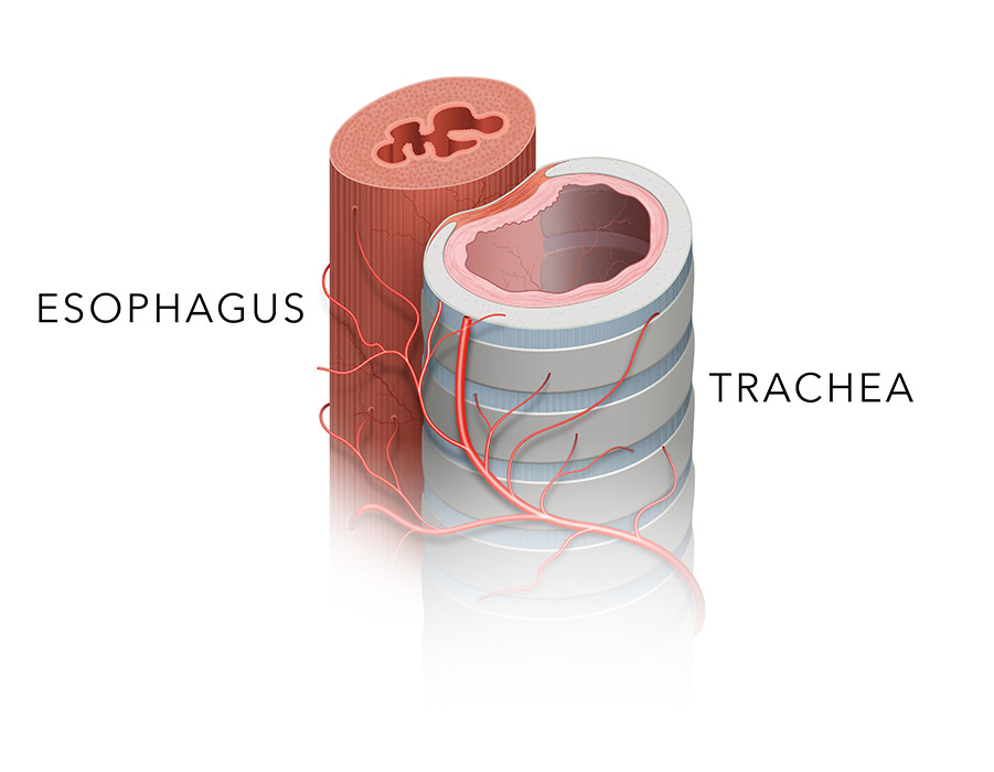
Our esophagus cell portfolio includes primary human cell types representing the major structural and functional components of the esophageal wall. These models support research in esophageal biology, epithelial barrier function, tissue remodeling, inflammation, microvascular physiology, and upper gastrointestinal diseases.
Available cell types include esophageal epithelial cells, fibroblasts, smooth muscle cells, and microvascular endothelial cells, enabling comprehensive studies of cellular interactions within the esophageal microenvironment and disease-related tissue responses.
Our Esophagus Cell Portfolio Highlights
Comprehensive Esophageal Cell Resources
Our collection encompasses key cellular components involved in esophageal structure, barrier maintenance, tissue repair, and vascular function.
-
Major Esophageal Cell Types
- Human esophageal epithelial cells
- Human esophageal fibroblasts
- Human esophageal smooth muscle cells
-
Microenvironment-Relevant Models
- Esophageal microvascular endothelial cells
- Models for epithelial-stromal interaction studies
- Suitable for tissue remodeling investigations
-
Primary Human Cell Resources
- Physiologically relevant human-derived models
- Available for a wide range of gastrointestinal research applications
Upper Gastrointestinal Research Support
We support studies focused on esophageal physiology, epithelial homeostasis, tissue injury, and disease-associated cellular responses.
-
Barrier Function Studies
Suitable for investigating epithelial integrity, permeability regulation, and responses to chemical, inflammatory, or mechanical stress.
-
Tissue Remodeling Research
Support for studies involving fibroblast activation, extracellular matrix deposition, wound repair, and fibrosis-associated processes.
-
Microvascular Biology Applications
Useful for evaluating angiogenesis, endothelial function, inflammatory cell recruitment, and vascular responses within esophageal tissues.
-
Multi-Cell Esophageal Models
Compatible cell types for co-culture systems that more closely replicate the esophageal microenvironment.
4
Core Esophageal
Cell Types
100 %
Mycoplasma-Free
Tested
24-72 h
Rapid
Delivery
Which esophageal cell type is most suitable for barrier function studies?
Human esophageal epithelial cells are the primary model for investigating epithelial integrity, permeability, cell junction regulation, and responses to environmental or inflammatory stimuli affecting the esophageal lining.
How are esophageal fibroblasts used in disease-related research?
Esophageal fibroblasts are commonly utilized to study extracellular matrix production, wound healing, tissue remodeling, and fibrosis-associated mechanisms that contribute to chronic esophageal disorders.
Can these cells be used to model the esophageal microenvironment?
Yes. Combining epithelial cells, fibroblasts, smooth muscle cells, and microvascular endothelial cells enables the development of co-culture systems that better mimic the cellular complexity of native esophageal tissues.
What applications are supported by esophageal microvascular endothelial cells?
These cells are valuable for studies of angiogenesis, vascular permeability, inflammatory signaling, endothelial activation, and vascular contributions to esophageal tissue injury and repair.
Are these cells suitable for studies of reflux-related esophageal injury?
Yes. Esophageal epithelial and stromal cell models are frequently used to investigate cellular responses to acid exposure, inflammatory mediators, tissue damage, and repair processes associated with reflux-related disorders.
Filters Clear all filters
Species
- Bovine (22)
- Cat (45)
- Chicken (11)
- Chinchilla (1)
- Dog (118)
- Fish (1)
- Fruitfly (1)
- Goat (46)
- Guinea Pig (8)
- Hamster (94)
- Horse (1)
- Human (800)
- Minipig (2)
- Monkey (129)
- Mouse (875)
- Pig (110)
- Rabbit (249)
- Rat (324)
- Sheep (2)
- Squirrel (1)
- Turkey (1)
Source
- Adipose (32)
- Adrenal Gland (11)
- Airway (8)
- Anus (3)
- Aorta (86)
- Artery (180)
- Bile Duct (9)
- Bladder (49)
- Blood (193)
- Bone (13)
- Bone Marrow (157)
- Brain (163)
- Breast (64)
- Bronchus (43)
- Cartilage (29)
- Cervix (5)
- Chorion (5)
- Choroid (9)
- Ciliary Body (1)
- Colon (63)
- Conjunctiva (9)
- Cord Blood (24)
- Cornea (27)
- Dental Pulp (4)
- Dermis (111)
- Diaphragm (3)
- Ear (12)
- Embryo (24)
- Endometrium (11)
- Epidermis (26)
- Epididymis (3)
- Esophagus (35)
- Eye (95)
- Foreskin (2)
- Gallbladder (4)
- Gingiva (20)
- Hair Follicle (15)
- Heart (72)
- Intestine (152)
- Iris (1)
- Kidney (151)
- Lens (4)
- Liver (117)
- Lung (193)
- Lymph Node (27)
- Mesentery (18)
- Nose (5)
- Olfactory Bulb (1)
- Oral Cavity (12)
- Ovary (72)
- Oviduct (7)
- Pancreas (68)
- Pancreatic Duct (3)
- Pancreatic Islet (11)
- Parathyroid Gland (4)
- Penis (7)
- Perineurium (1)
- Periodontal Ligament (5)
- Periodontium (25)
- Peripheral Blood (153)
- Peritoneal Cavity (14)
- Placenta (29)
- Prostate (62)
- Pudenda (2)
- Rectum (3)
- Retina (38)
- Salivary Gland (3)
- Sclera (3)
- Seminal Vesicle (1)
- Skeletal Muscle (36)
- Skin (155)
- Small Intestine (56)
- Spinal Cord (10)
- Spleen (76)
- Stomach (37)
- Synovial Fluid (2)
- Synovium (13)
- Tendon (8)
- Testis (15)
- Thymus (51)
- Thyroid (34)
- Tongue (6)
- Tonsil (3)
- Tooth (4)
- Trabecular Meshwork (3)
- Trachea (46)
- Umbilical Cord (29)
- Ureter (10)
- Urethra (3)
- Uterus (61)
- Vas Deferens (1)
- Vein (104)
Cell Type
- Adipocyte (4)
- Astrocyte (34)
- B Cell (30)
- Basal Cell (3)
- Basophil (1)
- Beta Cell (3)
- Cardiomyocyte (18)
- CD133+ Cell (6)
- CD34+ Cell (21)
- Cholangiocyte (9)
- Chondrocyte (19)
- Dendritic Cell (15)
- Endothelial Cell (688)
- Endothelial Progenitor Cell (7)
- Eosinophil (1)
- Epithelial Cell (516)
- Fibroblast (473)
- Glial Cell (58)
- Goblet Cell (1)
- Granule Cell (2)
- Granulocyte (12)
- Granulosa Cell (1)
- Hepatic Stellate Cell (9)
- Hepatocyte (22)
- Interstitial Cell (10)
- Keratinocyte (24)
- Keratocyte (3)
- Kupffer Cell (8)
- Leydig Cell (3)
- Lymphocyte (84)
- Macrophage (31)
- Mast Cell (3)
- Melanocyte (11)
- Meningeal Cell (5)
- Mesangial Cell (10)
- Mesothelial Cell (5)
- Microglia (7)
- Microvascular Cell (308)
- Monocyte (16)
- Mononuclear Cell (110)
- Myeloid Cell (2)
- Myoblast (5)
- Myofibroblast (3)
- Myosatellite Cell (2)
- Neuron (50)
- Neutrophil (10)
- NK Cell (11)
- Oligodendrocyte (3)
- Oligodendrocyte Progenitor Cell (4)
- Osteoblast (8)
- Osteoclast (2)
- Osteocyte (3)
- Pancreatic Stellate Cell (4)
- Pericyte (20)
- Podocyte (5)
- Preadipocyte (21)
- Progenitor Cell (15)
- Red Blood Cell (12)
- Retinal Ganglion Cell (3)
- Satellite Cell (2)
- Schwann Cell (4)
- Sebocyte (1)
- Sertoli Cell (5)
- Skeletal Muscle Cell (11)
- Smooth Muscle Cell (241)
- Spermatogonium (3)
- Stromal Cell (41)
- Synoviocyte (11)
- T Cell (39)
- Tenocyte (8)
- Trabecular Meshwork Cell (3)
- Trophoblast (4)
Disease
- Acute Lymphocytic Leukemia (ALL) (15)
- Acute Myeloid Leukemia (AML) (13)
- Amyotrophic Lateral Sclerosis (ALS) (4)
- Aplastic Anemia (AA) (1)
- Arteriovenous Malformation (AVM) (1)
- Asthma (5)
- Astrocytoma (2)
- Autoimmune Hemolytic Anemia (AIHA) (1)
- Autoimmune Lymphoproliferative Syndrome (ALPS) (1)
- Breast Cancer (8)
- Cancer (144)
- Cervical Cancer (2)
- Chronic Lymphocytic Leukemia (CLL) (19)
- Chronic Myeloid Leukemia (CML) (14)
- Chronic Obstructive Pulmonary Disease (COPD) (6)
- Colon Cancer (9)
- Crohn's Disease (3)
- Cystic Fibrosis (CF) (7)
- Diabetes (110)
- Diabetes Type 1 (16)
- Diabetes Type 2 (18)
- Diffuse Large B-Cell Lymphoma (4)
- Dilated Cardiomyopathy (DCM) (1)
- Duchenne Muscular Dystrophy (DMD) (5)
- Essential Thrombocythemia (ET) (1)
- Glioblastoma (3)
- Guillain-Barre Syndrome (GBS) (1)
- Hypertension (27)
- Idiopathic Thrombocytopenic Purpura (ITP) (1)
- Inflammatory Bowel Disease (IBD) (5)
- Iron-Deficiency Anemia (1)
- Kidney Cancer (3)
- Legg–Calvé–Perthes Disease (LCPD) (2)
- Leukopenia (1)
- Liver Cancer (3)
- Lung Cancer (12)
- Mantle Cell Lymphoma (MCL) (8)
- Melanoma (2)
- Mucopolysaccharidosis (2)
- Multiple Myeloma (MM) (12)
- Multiple Sclerosis (MS) (3)
- Muscular Dystrophy (MD) (1)
- Myelodysplastic Syndrome (MDS) (3)
- Neurofibromatosis (NF) (3)
- Non-Hodgkin Lymphoma (NHL) (10)
- Normal (2466)
- Osteoarthritis (OA) (5)
- Ovarian Cancer (6)
- Pancreatic Cancer (3)
- Pancytopenia (1)
- Parkinson's Disease (PD) (2)
- Plasmacytoma (1)
- Polycythemia (1)
- Prostate Cancer (6)
- Psoriasis (4)
- Rheumatoid Arthritis (RA) (7)
- Robertsonian Translocation (ROB) (1)
- Sickle Cell Anemia (2)
- Systemic Lupus Erythematosus (SLE) (4)
- Thrombocytopenia (1)
- Transverse Myelitis (TM) (1)
- Ulcerative Colitis (UC) (2)
- Waldenström Macroglobulinemia (WM) (2)

Description: Smooth muscle is responsible for the contractility of hollow organs, such as blood vessels, the ...

Description: The HEF cells from Creative Bioarray are isolated from human esophageal tissue. HEF are ...
Description: The human esophagus is lined by a non-keratinizing, moist stratified squamous epithelium whose ...
Description: HEMEC from Creative Bioarray are isolated from human esophageal tissue. HEMEC are cryopreserved at ...