Monosodium Iodoacetate (MIA)-Induced Osteoarthritis (OA) Model
Creative Bioarray provides a monosodium iodoacetate (MIA)-induced osteoarthritis (OA) model for our clients to study the disease mechanisms or evaluate the efficacy of drug candidates. With our services, you can obtain reliable and accurate results, providing valuable insights into drug development.
OA is the most common form of arthritis among middle-aged and elderly individuals. Its etiology is complex and involves multiple factors, with the underlying pathogenic mechanisms remaining elusive. While the progression of the disease is typically slow, it can result in significant pain and disability, negatively impacting the quality of life. Given the challenges in studying OA in humans, animal models have become crucial in assessing the development of the disease and evaluating innovative treatment strategies.
Currently, the most utilized type of chemical induction is MIA, which has an inhibitory activity of glyceraldehyde-3-phosphate dehydrogenase glycolysis and induces the death of chondrocytes. In both rodents and non-rodents, the intra-articular injection causes chondrocyte destruction. When administered in rats, it causes cartilage lesions with loss of proteoglycan matrix and functional alterations with stiffness that are analogous to those seen in human osteoarthritis.
Our Monosodium Iodoacetate (MIA)-Induced Osteoarthritis (OA) Model
- Available Animal
Rat - Modeling Method
After anesthetization, rats are given a single intra-articular injection of MIA through the infrapatellar ligament of the right knee. - Endpoints
- Behavioral test
- Histology analysis (knee joint): H&E staining, Safranin-O/Fast Green staining
- Mankin score
- Serum analysis: cytokine, SOD, MDA, etc.
- Body weight
- Other customized endpoints
Example Data
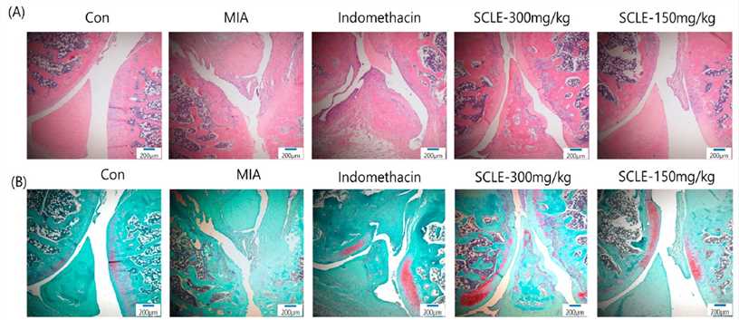 Fig. 1 Histopathological features of knee joint tissues in monosodium iodoacetate (MIA)-induced osteoarthritis (OA) rats. Knee joint tissues histopathological changes with (A) H&E and (B) Safranin-O/Fast Green
Meanwhile, we also provide other OA models that maybe you are interested in:
- Papain-Induced Osteoarthritis (OA) Model
- Medial Meniscal Tear (MMT)-Induced Osteoarthritis (OA) Model
- Anterior Cruciate Ligament Transection (ACLT)-Induced Osteoarthritis (OA) Model
Quotation and Ordering
With years of experience in disease models, the scientists at Creative Bioarray are eager to collaborate with our clients to create a customized design tailored to your unique needs. If you are interested in our services, please feel free to contact us at any time or submit an inquiry to us directly.
References
- Lee, Y.M., et al. Anti-inflammatory and analgesic effects of Schisandra chinensis leaf extracts and monosodium iodoacetate-induced osteoarthritis in rats and acetic acid-induced writhing in mice. Nutrients, 2022, 14(7): 1356.
- Longo, U.G., et al. Induced models of osteoarthritis in animal models: a systematic review. Biology, 2023, 12(2): 283.
For research use only. Not for any other purpose.
Disease Models
- Oncology Models
-
Inflammation & Autoimmune Disease Models
- Rheumatoid Arthritis Models
- Glomerulonephritis Models
- Multiple Sclerosis (MS) Models
- Ocular Inflammation Models
- Sjögren's Syndrome Model
- LPS-induced Acute Lung Injury Model
- Peritonitis Models
- Passive Cutaneous Anaphylaxis Model
- Delayed-Type Hypersensitivity (DTH) Models
- Inflammatory Bowel Disease Models
- Systemic Lupus Erythematosus Animal Models
- Oral Mucositis Model
- Asthma Model
- Sepsis Model
- Psoriasis Model
- Atopic Dermatitis (AD) Model
- Scleroderma Model
- Gouty Arthritis Model
- Carrageenan-Induced Air Pouch Synovitis Model
- Carrageenan-Induced Paw Edema Model
- Experimental Autoimmune Myasthenia Gravis (EAMG) Model
- Graft-versus-host Disease (GvHD) Models
-
Cardiovascular Disease Models
- Surgical Models
- Animal Models of Hypertension
- Venous Thrombosis Model
- Atherosclerosis model
- Cardiac Arrhythmia Model
- Hyperlipoidemia Model
- Doxorubicin-induced Heart Failure Model
- Isoproterenol-induced Heart Failure Model
- Arterial Thrombosis Model
- Pulmonary Arterial Hypertension (PAH) Models
- Heart Failure with Preserved Ejection Fraction (HFpEF) Model
- Cardio-Renal-Metabolic (CKM) Syndrome Model
-
Neurological Disease Models
- Alzheimer's Disease Modeling and Assays
- Ischemic Stroke Models
- Acute Spinal Cord Injury (ASCI) Model
- Traumatic Brain Injury (TBI) Model
- Hypoxic-Ischemic Encephalopathy (HIE) Model
- Tourette Syndrome (TS) Model
- Amyotrophic Lateral Sclerosis (ALS) Model
- Huntington's Disease (HD) Model
- Intracerebral hemorrhage (ICH) Models
- Schizophrenia Model
- Depression Models
- Pain Models
-
Metabolic Disease Models
- Type 1 Diabetes Mellitus Model
- Type 2 Diabetes Mellitus Model
- Animal Model of Hyperuricemia
-
Nonalcoholic Fatty Liver Disease Model
- High-Fat Diet-Induced Nonalcoholic Fatty Liver Disease (NAFLD) Model
- Methionine and Choline Deficient (MCD) Diet-Induced Nonalcoholic Fatty Liver Disease (NAFLD) Model
- Gubra-Amylin NASH (GAN) Diet-Induced Nonalcoholic Fatty Liver Disease (NAFLD) Model
- Streptozotocin (STZ) Induced Nonalcoholic Fatty Liver Disease (NAFLD) Model
- High Fat Diet-Induced Obesity Model
- Diabetic Foot Ulcer (DFU) Model
- Liver Disease Models
- Rare Disease Models
- Respiratory Disease Models
- Digestive Disease Models
-
Urology Disease Models
- Cisplatin-induced Nephrotoxicity Model
- Unilateral Ureteral Obstruction Model
- 5/6 Nephrectomy Model
- Renal Ischemia-Reperfusion Injury (RIRI) Model
- Diabetic Nephropathy (DN) Models
- Passive Heymann Nephritis (PHN) Model
- Adenine-Induced Chronic Kidney Disease (CKD) Model
- Kidney Stone Model
- Doxorubicin-Induced Nephropathy Model
- Orthotopic Kidney Transplantation Model
- Benign Prostatic Hyperplasia (BPH) Model
- Peritoneal Fibrosis Model
- Orthopedic Disease Models
- Ocular Disease Models
- Infectious Disease Models
- Skin Disease Models
- Otology Disease Models