
Unilateral Ureteral Obstruction Model
A validated, reproducible platform for preclinical evaluation of tubulointerstitial fibrosis therapies. From surgical execution to IND-enabling data packages.
The UUO Model in Preclinical Research
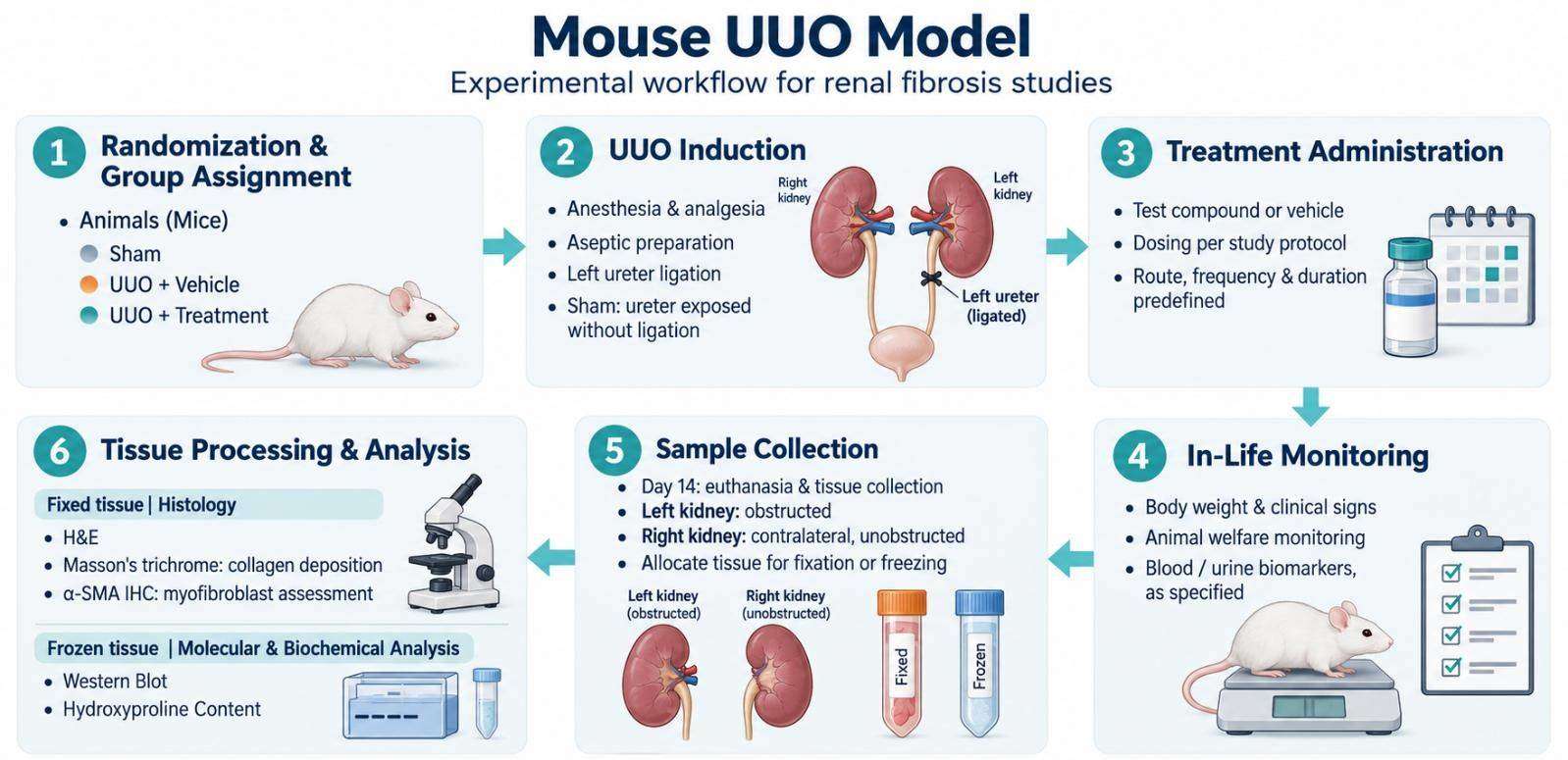
Unilateral Ureteral Obstruction (UUO) is the most widely validated and reproducible animal model of renal tubulointerstitial fibrosis. By surgically ligating one ureter, the model induces progressive obstructive nephropathy characterized by tubular atrophy, inflammatory cell infiltration, myofibroblast activation, and extracellular matrix (ECM) accumulation — pathological hallmarks that closely mirror human chronic kidney disease (CKD) progression.
As a specialized Preclinical CRO, we provide standardized, GLP-compliant UUO model services to accelerate your nephrology and fibrosis therapeutic pipeline from lead validation to IND-enabling studies.
Why the UUO Model?
The UUO model offers distinct advantages for preclinical pharmacology:
- Rapid Fibrosis Onset: Significant tubulointerstitial fibrosis develops within 14 days post-obstruction, enabling fast turnaround for compound screening.
- High Reproducibility: Surgical technique is well-established; inter-animal variability is low when performed by trained veterinary surgeons.
- Mechanistic Clarity: The model isolates tubulointerstitial injury from hemodynamic confounders (unlike diabetic or hypertensive nephropathy models), allowing direct assessment of antifibrotic mechanisms.
- Translatability: Molecular pathways (TGF-β1/Smad, Notch, Wnt/β-catenin, inflammation) and histopathological outcomes are highly conserved across rodents and humans.
- Bilateral Readout Control: The contralateral, unobstructed kidney serves as an internal within-subject control, reducing biological variance.
Model Specifications & Surgical Protocol
Animal strains
- C57BL/6J mouse
Surgical procedure
- Anesthesia: Isoflurane inhalation (2–3% maintenance) with continuous physiological monitoring.
- Ureteral Ligation: A midline abdominal incision exposes the left ureter; double ligation with 4-0 silk suture is performed at the level of the lower pole of the kidney, followed by ureteral transection between ligatures to prevent retrograde infection.
- Sham Control: Identical surgical exposure without ureteral ligation.
Standard study durations
- Acute Injury Phase: 3–5 days post-obstruction: tubular dilation, inflammatory influx
- Fibrogenic Phase: 7–10 days: myofibroblast activation, ECM synthesis
- Established Fibrosis: 14–21 days: advanced collagen deposition, tubular atrophy
Comprehensive Endpoint Portfolio
We offer comprehensive, customizable endpoint analysis to address efficacy, mechanism of action, and safety:
1. Histopathology & Histomorphometry
- H&E staining for tubular injury scoring (tubular dilatation, cast formation, atrophy)
- Masson's Trichrome staining for collagen deposition
- Immunohistochemistry (IHC) for marker expression
2. Fibrosis Biomarkers
- α-SMA (α-smooth muscle actin) IHC, Western blot, or qPCR
- Collagen type I / III mRNA and protein
- Hydroxyproline assay for total collagen content quantification
3. Kidney Injury & Function
- Kidney weight / body weight ratio
- Serum BUN and creatinine (optional; note that UUO does not cause severe systemic uremia)
- Urinary kidney injury molecule-1 (KIM-1, ureteral obstruction)
- NGAL (neutrophil gelatinase-associated lipocalin)
- KIM-1 mRNA/protein expression in renal cortex
4. In Situ & Molecular Localization
- In situ hybridization / RNAscope for target genes
- Immunofluorescence (IF) co-staining for α-SMA and podocyte or tubular markers (optional)
Therapeutic Applications & Indications
Our UUO platform supports preclinical evaluation across diverse therapeutic modalities:
1. Antifibrotic Small Molecules
- TGF-β receptor kinase inhibitors
- LOX/LOXL2 inhibitors (collagen cross-linking blockers)
- NOX inhibitors (oxidative stress attenuation)
2. Biologics & Antibody Therapeutics
- Anti-CTGF antibodies
- Anti-TGF-β monoclonal antibodies
- Anti-inflammatory biologics targeting IL-1β or TNF-α pathways
3. Gene & Cell Therapy
- siRNA / antisense oligonucleotides targeting profibrotic genes
- Mesenchymal stem cell (MSC) therapy efficacy and biodistribution
- AAV-mediated gene delivery studies
4. Natural Products & Nutraceuticals
- Standardized herbal extracts with renoprotective claims
- Metabolite-based interventions
Our CRO Capabilities & Quality Assurance

Surgical Expertise
- Our veterinary surgical team performs >500 renal surgeries annually with a complication rate <3% and mortality <2%.
- All procedures are conducted in dedicated surgical suites with aseptic technique.

Histopathology Core
- Board-certified veterinary pathologists with nephropathology specialization
- Blind scoring protocols to eliminate bias
Custom Study Design
- Dose-response and time-course optimization
- Combination therapy studies (e.g., ACE inhibitor + antifibrotic agent)
- Deobstruction/reversal paradigms to test disease-modifying vs. preventive efficacy
Representative Data & Case Study
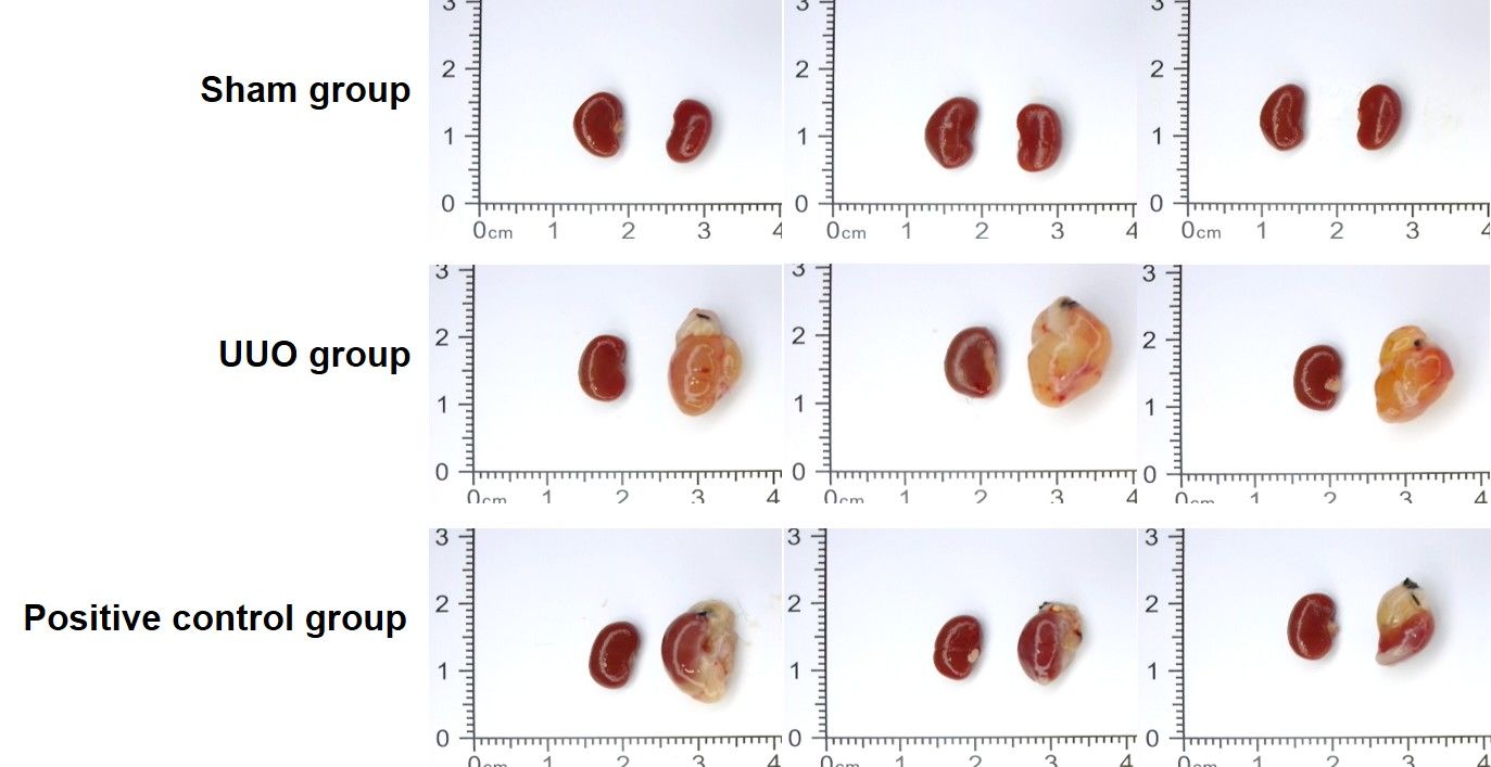 Figure 1. Representative gross images of kidneys from mice subjected to unilateral ureteral obstruction (UUO).
The UUO model induced visible renal morphological changes compared with Sham-operated animals, while positive control treatment showed improvement of UUO-associated kidney alterations.
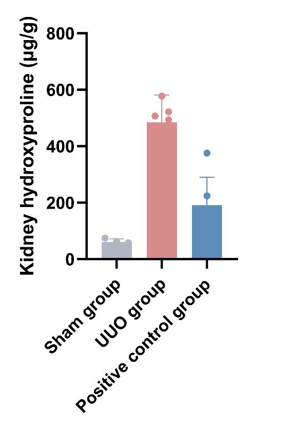 Figure 2. Quantification of kidney hydroxyproline content in the UUO model.
UUO induction significantly increased renal hydroxyproline levels compared with the Sham group, indicating enhanced collagen deposition. Positive control treatment partially attenuated UUO-induced fibrosis-associated changes.
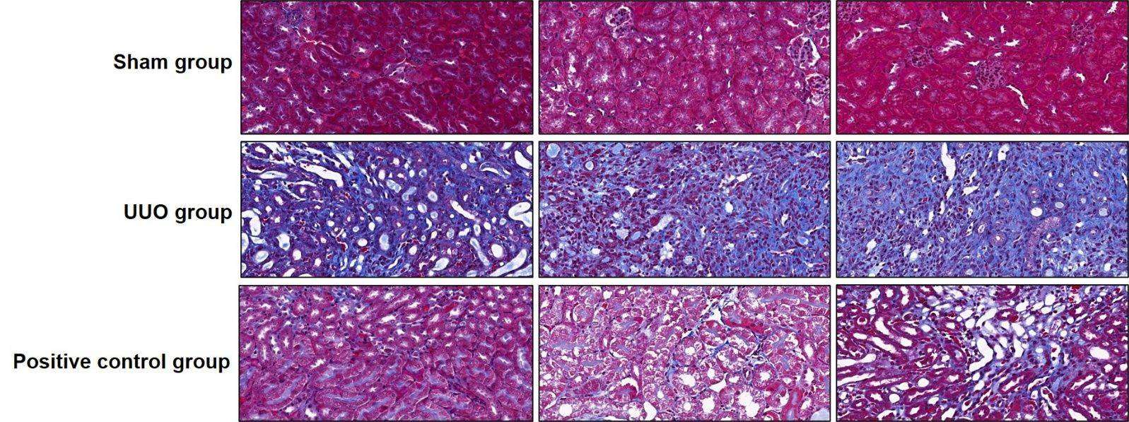 Figure 3. Masson's trichrome staining of kidney tissues in the UUO model.
Masson staining revealed increased collagen deposition and renal fibrosis following UUO induction compared with Sham controls. Positive control treatment reduced UUO-associated fibrotic changes.
FAQ
How does the UUO model differ from the 5/6 nephrectomy or diabetic nephropathy models?
The UUO model induces rapid tubulointerstitial fibrosis through mechanical ureteral obstruction, making it particularly suitable for investigating renal fibrosis mechanisms and evaluating potential antifibrotic therapies. In contrast, the 5/6 nephrectomy model is commonly used to study progressive chronic kidney disease (CKD) following nephron loss, while diabetic nephropathy models focus on diabetes-associated kidney injury. For studies targeting glomerular injury or specific causes of CKD, alternative models, such as 5/6 nephrectomy, diabetic nephropathy, or adriamycin-induced nephropathy, may be more appropriate.
What is the minimum group size for statistically robust fibrosis quantification?
A group size of 8–10 animals is commonly considered for UUO fibrosis studies. However, the optimal sample size depends on the study design, expected treatment effects, and variability of the selected endpoints. We can provide recommendations based on specific study requirements.
Ready to Advance Your Renal Fibrosis Program?
Contact our scientific team to discuss your specific therapeutic target, preferred endpoints, and study timeline. We provide no-obligation protocol consultation and feasibility assessments within 24 hours.