
CAL-27
Cat.No.: CSC-C0478
Species: Homo sapiens (Human)
Source: Oral Cavity; Tongue
Morphology: Epithelial
Culture Properties: monolayer
- Specification
- Background
- Scientific Data
- Q & A
- Customer Review
CSF1PO: 10,12
D13S317: 10,11
D16S539: 11,12
D18S51: 13
D21S11: 28,29
D3S1358: 16
D5S818: 11,12
D7S820: 10
D8S1179: 13,15
FGA: 25
Penta D: 9,10
Penta E: 7
TH01: 6,9,3
TPOX: 8
vWA: 14,17
Immunology: cytokeratin +, cytokeratin-7 -, cytokeratin-8 +, cytokeratin-17 +, cytokeratin-18 +, cytokeratin-19 -, desmin -, endothel -, EpCAM +, GFAP -, neurofilament -
CAL-27 Cell Line is a human tongue squamous cell carcinoma cell line established from a poorly differentiated primary tumor of the tongue obtained from a 56-year-old male patient. It is commonly used as an in vitro model for studying head and neck squamous cell carcinoma (HNSCC) and exhibits stable growth, genetic reproducibility, and a well-defined phenotype representing the molecular and pathological features of oral cancers. Morphologically, CAL-27 cells are epithelial-like, polygonal, and exhibit strong adherence to the culture surface, forming tight, carcinoma-derived colonies.
Functionally, CAL-27 cells demonstrate high proliferative and invasive potential, express cytokeratins characteristic of squamous epithelium, and carry mutations in key oncogenes and tumor suppressor genes such as TP53, reflecting HNSCC's molecular alterations. These features make CAL-27 useful for studying tumor progression, metastasis, and epithelial-mesenchymal transition (EMT) mechanisms. They are frequently used in pharmacological and therapeutic studies, including chemoresistance, radiotherapy response, and targeted therapy evaluation. CAL-27 cells also facilitate research on tumor-microenvironment interactions and the regulation of genes involved in oral cancer pathogenesis.

Molecular correlates for HPV-negative head and neck cancer engraftment prognosticate patient outcomes
Background
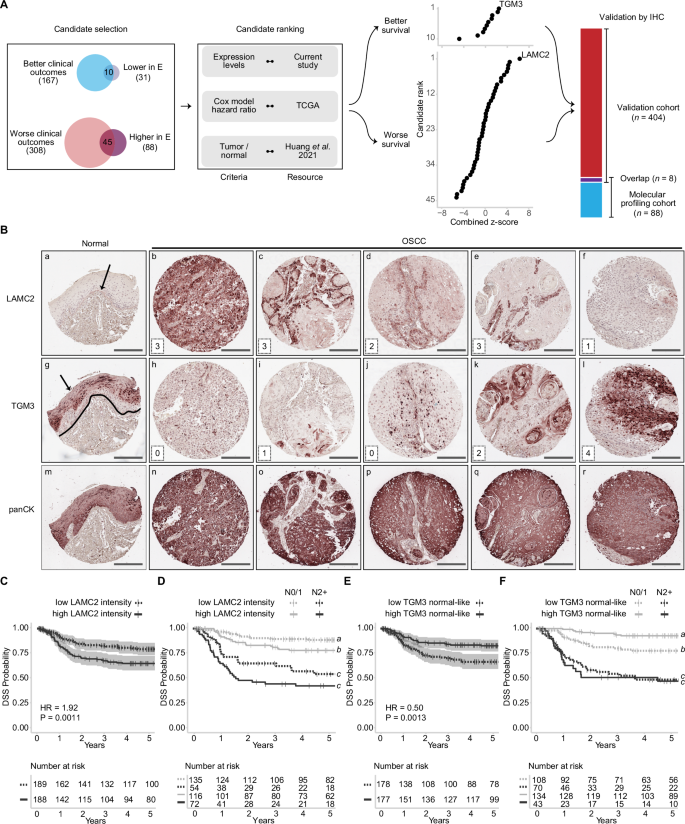
HPV-negative head and neck squamous cell carcinoma (HNSCC) needs better risk stratification due to treatment toxicity. Lymph node involvement is a key prognostic factor, and patient-derived xenograft engraftment predicts poor outcomes but is impractical clinically. The authors performed transcriptomic and proteomic profiling of 88 HPV-negative HNSCC tumors and found that the relationship between engraftment and clinical outcomes is recapitulated by molecular phenotype. They identified LAMC2 and TGM3 as candidate prognostic biomarkers and validated them in an independent cohort of 404 HPV-negative HNSCC patients, showing improved outcome prediction beyond nodal status alone.
Key Findings
Engraftment of HPV-negative HNSCC tumors in mice was significantly associated with worse disease-specific survival and provided risk stratification beyond N category. Molecular profiling linked engraftment to partial epithelial-mesenchymal transition (p-EMT), extracellular matrix reorganization, diminished immune signaling, and fibroblast-epithelial cell communication. LAMC2 and TGM3 were identified as top candidate biomarkers: high LAMC2 intensity correlated with worse outcomes, while normal-like TGM3 staining correlated with better outcomes. In an independent cohort of 404 HNSCC patients, immunohistochemical scoring of LAMC2 and TGM3 validated these associations and significantly stratified outcomes beyond nodal status. Combining LAMC2 and TGM3 improved prognostication within N0 patients, identifying subsets with markedly different failure rates and survival, and suggested potential utility for elective neck dissection decisions and risk-tailored treatment planning.
Contribution
Creative Bioarray provided GFP-labeled Cal33 and Cal27 head and neck squamous cell carcinoma cell lines used in co-culture spheroid experiments with patient-derived cancer-associated fibroblasts. These reagents enabled the authors to assess fibroblast-epithelial crosstalk and p-EMT induction, supporting mechanistic links between CAF signaling and engraftment-associated molecular phenotypes.
Effect of Honokiol-Loaded Titanium Dioxide Nanotube Drug Delivery System on CAL-27 Cells
Tongue cancer is the most common type of oral cancer with a low postoperative prognosis. Honokiol (HNK), a compound extracted from Houpu, has been shown to have anti-tumor effects. Titanium dioxide nanotubes (TNTs) can be used as drug carriers. The purpose of this experiment was to study the anti-tumor effect of HNK-loaded TNTs (HNK-TNTs) on CAL-27 cells.
The results of CCK-8 experiment showed that compared with the control group, the proliferation rate of CAL-27 cells in group HNK and HNK-TNTs was negatively correlated with the time of drug action, and the proliferation rate in group HNK-TNTs was lower than that in group HNK (Fig. 1), suggesting that HNK-TNTs has a better effect on CAL-27 cells proliferation inhibition than HNK. As shown in this scratching experiment results, after 12 and 24 h of cell culture, the migration ability of CAL-27 cells in group HNK-TNTs was significantly lower than that in group control and group HNK (Fig. 2A), and the difference was statistically significant (Fig. 2B). It was indicated that HNK-TNTs has a more significant inhibitory effect on the cell migration.


Bacterial Antigens Reduced the Inhibition Effect of Capsaicin on Cal 27 Oral Cancer Cell Proliferation
Oral cancer is a significant global health issue with high incidence and low survival rates. The oral cavity contains biofilms, including dental plaques, which harbor bacterial antigens like LPS and LTA from Gram-negative and Gram-positive bacteria. These antigens can stimulate cancer cell growth, while capsaicin has been reported to counteract this effect. Chakraborty et al. aim to evaluate the efficacy of capsaicin in treating oral cancer, particularly in the presence of LPS, LTA, or both.
Titration of capsaicin found that the maximum clinically relevant concentration that could be used without adversely affecting normal cells was 150 µM. Capsaicin at 150 µM produced maximum inhibition of oral cancer cell Cal 27 (Fig. 3a), but a small effect on cell viability of normal oral cell OKF6 (Fig. 4b). Therefore, they could not increase the concentration of capsaicin in the study. Within one hour of application, capsaicin at 150 µM reduced 60% of Cal 27 metabolism (Fig. 3b). The level of inhibition remained steady for up to 24 h (Fig. 3b). There was no significant change in normal oral cell OKF6 metabolism (Fig. 4a) and a small effect on cell viability (Fig. 4b) when treated with 150 µM capsaicin or stimulated with bacterial antigens.


Ask a Question
Write your own review
- You May Also Need

Description: Human cell line derived from oral squamous cell carcinoma occurred in 69-yo, male patient. HLA-A 24/.
Description: Human cell line derived from metastasis of cancer occurred in oral cavity, the same patient as T3M-1 Clone2 and T3M-1 Cl-10.
Description: G-CSF and IL-1 producing oral squamous cell carcinoma, the same patient as T3M-1 Clone2 and CJM.
Description: Established from the surgically removed fragment of a tongue lesion from a 69-year-old man with moderately differentiated squamous cell carcinoma of the tongue in 1983 (prior to therapy)
- Adipose Tissue-Derived Stem Cells
- Human Neurons
- Mouse Probe
- Whole Chromosome Painting Probes
- Hepatic Cells
- Renal Cells
- In Vitro ADME Kits
- Tissue Microarray
- Tissue Blocks
- Tissue Sections
- FFPE Cell Pellet
- Probe
- Centromere Probes
- Telomere Probes
- Satellite Enumeration Probes
- Subtelomere Specific Probes
- Bacterial Probes
- ISH/FISH Probes
- Exosome Isolation Kit
- Human Adult Stem Cells
- Mouse Stem Cells
- iPSCs
- Mouse Embryonic Stem Cells
- iPSC Differentiation Kits
- Mesenchymal Stem Cells
- Immortalized Human Cells
- Immortalized Murine Cells
- Cell Immortalization Kit
- Adipose Cells
- Cardiac Cells
- Dermal Cells
- Epidermal Cells
- Peripheral Blood Mononuclear Cells
- Umbilical Cord Cells
- Monkey Primary Cells
- Mouse Primary Cells
- Breast Tumor Cells
- Colorectal Tumor Cells
- Esophageal Tumor Cells
- Lung Tumor Cells
- Leukemia/Lymphoma/Myeloma Cells
- Ovarian Tumor Cells
- Pancreatic Tumor Cells
- Mouse Tumor Cells