Wound Healing

A better understanding of the wound healing process leads to generating better products that aid in healing hard-to-heal wounds such as chronic wounds. Cell migration during the wound healing process is an important step of a complete healing. In vivo, the wound healing process includes different cell types (macrophages, fibroblasts, and keratinocytes) coming in and out of the wound bed at different time points. Mimicking the in vivo milieu with an in vitro 3D model will assist wound healing scientists to better understand the wound healing or re-epithelialization process and aid companies in generating better products of helping with hard-to-heal acute wounds and chronic wounds.
Creative Bioarray provides in vitro 3D skin tissue models and evaluation assays to support cosmetic or dermatological industries in generating products of helping with wound healing or re-epithelialization.
Your Needs
- You are looking for a skin model to perform in vitro wound healing testing?
- You wish to screen ingredients, and characterize the efficacy or toxicity or safety of any products or chemicals of wound healing?
- You'd like to find a customized in vitro testing service of wound healing?
Our Capability
In vitro 3D skin models available
- Normal human epidermal keratinocytes (NHEK)
- Normal human dermal fibroblasts (NHDF)
- Full thickness skin model (FTSK)
- Reconstructed human epidermis (RHE)
- Ex vivo skin explants
Screening assays available
- Evaluating effects of active compounds related to wound healing
- Evaluating effects of different formulations
Endpoints
- Cell proliferation
- Migration ("scratch" assay, Boyden chambers)
- Invasion (Boyden chambers)
- Sequential expression of tissue repair markers(cytokine secretion, protease activities).
- Inflammatory response
- Neovascularization and neuritogenesis
- Extracellular matrix synthesis, fibrosis and myofibroblast interaction
- Dermal remodeling and epidermal differentiation
Techniques
- qPCR, qPCRarray, RT-PCR
- Epidermal separation
- Immunofluorescence
- ELISA
- RNA extraction
- Protein extraction
- Macroscopy
Assay Examples
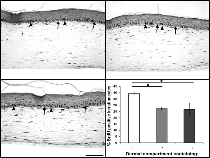 Fig1. Increased basal keratinocyte proliferation in 3D FTSK model. Representative images of tissue sections stained
for BrdU incorporation are shown (arrowheads mark examples of BrdU-stained nuclei, and arrows mark
examples of BrdU-negative nuclei). Quantification of mean percentage of basal keratinocytes positive for BrdU±SD.
FTSK, full thickness skin model.
Fig1. Increased basal keratinocyte proliferation in 3D FTSK model. Representative images of tissue sections stained
for BrdU incorporation are shown (arrowheads mark examples of BrdU-stained nuclei, and arrows mark
examples of BrdU-negative nuclei). Quantification of mean percentage of basal keratinocytes positive for BrdU±SD.
FTSK, full thickness skin model.
Related Products and Services
Choose our models to perform screening assays in house, or choose our assays and services directly!!
- Normal human epidermal keratinocytes (NHEK)
- Normal human dermal fibroblasts (NHDF)
- Full thickness skin model (FTSK)
- Reconstructed human epidermis (RHE)
- Ex vivo skin explants
- Compound screening service
Our customer service representatives are available 24hr a day! We thank you for choosing Creative Bioarray services!
In vitro Skin Models:
Explore Other Options