Ischemic Stroke Models
If you are looking for reliable and high-quality ischemic stroke models, Creative Bioarray has got you covered. Our models are well-established and trusted by clients around the world. With our models, you can study the mechanisms and injury caused by ischemic stroke and evaluate the efficacy of your test compounds on ischemic stroke. Our models are designed to help you gain deeper insights into the disease and develop effective treatments that can make a difference for patients. So why wait? Contact us now to learn more about our ischemic stroke models and start your research journey with confidence!
Stroke, a major global health issue, is the second leading cause of death and the leading cause of long-term disability worldwide. Stoke is divided into two major categories: ischemic and hemorrhagic. Ischemic stroke, the more common type, accounts for approximately 85% of all strokes. This condition occurs when there is a disruption in the blood supply to the brain, resulting in insufficient blood flow to meet the metabolic demands of the brain tissue. Hemorrhagic stroke, on the other hand, accounts for the remaining 15% of stroke. It occurs when a blood vessel within the brain ruptures, leading to bleeding into the brain tissue.
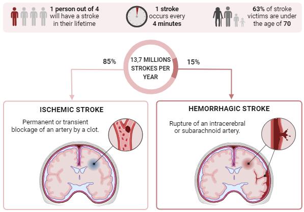 Fig. 1 Epidemiology and definitions of ischemic and hemorrhagic strokes
Our Animal Models of Ischemic Stroke
In the field of stroke research, numerous well-established ischemic stroke models exist, encompassing both global and focal ischemia models. As a leading research partner with extensive expertise in pharmacology and efficacy studies for various neurological disorders, Creative Bioarray is equipped to evaluate potential therapeutics using the following models:
Example Data
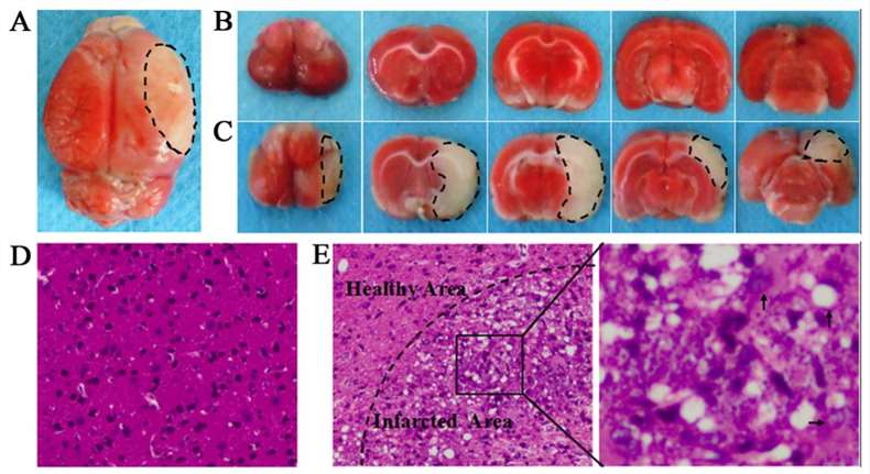 Fig. 2 (A) The complete brain of middle cerebral artery occlusion (MCAO) models. (B) Serial coronal slices of sham-operated rats. (C) Consecutive coronal slices derived from MCAO models. 2,3,5-Triphenyltetrazolium chloride (TTC) staining showed red healthy zones and pale infarcted regions; Cerebral infarct volume was ~32.1±1.8% in saline group. (D) H&E staining of sham-operated animals revealed intact brain tissues, with uniform, round or oval neurons and pink cytoplasm with abundant and clear nucleus. No inflammatory cells were detected. (E) H&E staining of MCAO models revealed lesions in the brain tissues, with diminished numbers of neurons and chaotic neuronal configuration.
Quotation and Ordering
Creative Bioarray establishes flexibly tailored study protocols that are designed to fit your specific needs. Our experienced scientists are well-versed in rapidly validating models from the literature, ensuring that our clients receive accurate data and results. If the model you require is not presented on our website, please do not hesitate to contact us. We would be more than happy to work with you and tailor our program to meet your specific needs.
Reference
- Zhang, X. et al. Intravenous administration of DPSCs and BDNF improves neurological performance in rats with focal cerebral ischemia. International Journal of Molecular Medicine, 2018, 41(6): 3185-3194.