Pristane Induced Model
Pristane is an isoprenoid alkane found at high concentration in plants, shark liver and mineral oil. Pristane induced model using B6 or Balb/c mice usually recapitulate clinical features of systemic lupus erythematosus, including high expression of anti-ribonucleic acid protein, anti-DNA and anti-histone autoantibodies and immune complex deposition. In addition, pristane-induced lupus is more pronounced in females than that in male. Creative Bioarray focuses on drug research and development services and helps customers test the efficacy of drug candidates targeting SLE and elucidate the pathogenesis by this model.
Our capabilities
- We detect autoantibodies in animal serum by ELISA, Immunofluorescence, etc.
- We evaluate various cytokines through IHC, ELISA, etc.
- We identify immunocyte subsets in spleens by flow cytometry.
Assays available
- PK/PD blood collections
- Quantitative detection of Anti-dsDNA and total IgG by ELISA
- Clinical chemistry analysis
- Histopathological evaluation
- Cytokine/chemokine analysis
- Flow cytometry in lymph nodes, spleen or bone marrow
- Organ weights (kidneys, spleen, lymph nodes)
- Survival rate
With extensive experience in the field of SLE, we are confident to help you to overcome any upcoming challenges. Our experts are fully capable of customizing our protocols and assays to meet your specific needs. With our help, we wish to facilitate your research with high efficiency.
Study examples
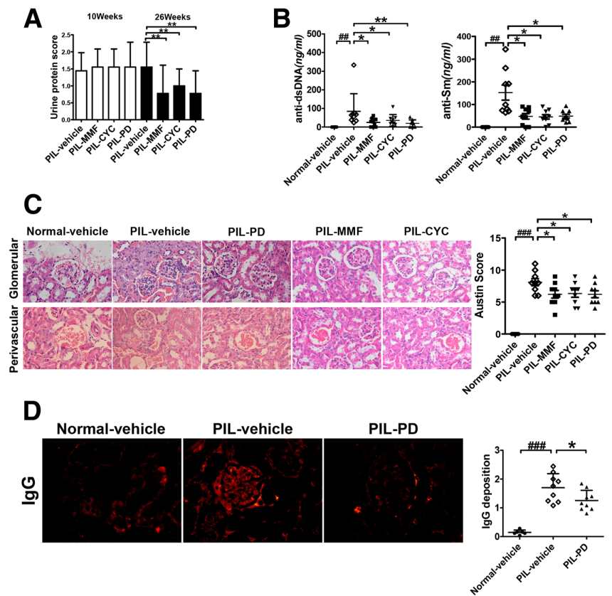 Figure. 1. PD ameliorated lupus manifestations in the kidneys in Pristane induced lupus mouse model (PIL mouse model). PIL mice were treated with vehicle or with PD (45 mg/kg, daily), CYC (1.8 mg/mouse, weekly), MMF (100 mg/kg, daily) for 16 weeks. A The proteinuria was assessed as described in Materials and Methods. B The levels of anti-dsDNA antibodies and anti-Sm antibodies were examined by ELISA. C On the left, representative H&E staining of glomerular and renal vascular lesions in kidneys was shown (× 400). On the right, Austin scores of kidneys were shown. D On the left, the glomeruli were stained for IgG deposition and the representative staining images were shown (× 400). On the right, 25 glomeruli were analyzed and the average score was calculated for each kidney as described in Materials and Methods.
Quotation and ordering
If you have any special needs or questions regarding our services, please feel free to contact us. We look forward to cooperating with you in the future.
Reference
Liao P et al. Polydatin effectively attenuates disease activity in lupus-prone mouse models by blocking ROS-mediated NET formation[J]. Arthritis research & therapy, 2018, 20(1).
For research use only. Not for any other purpose.
Disease Models
- Oncology Models
-
Inflammation & Autoimmune Disease Models
- Rheumatoid Arthritis Models
- Glomerulonephritis Models
- Multiple Sclerosis (MS) Models
- Ocular Inflammation Models
- Sjögren's Syndrome Model
- LPS-induced Acute Lung Injury Model
- Peritonitis Models
- Passive Cutaneous Anaphylaxis Model
- Delayed-Type Hypersensitivity (DTH) Models
- Inflammatory Bowel Disease Models
- Systemic Lupus Erythematosus Animal Models
- Oral Mucositis Model
- Asthma Model
- Sepsis Model
- Psoriasis Model
- Atopic Dermatitis (AD) Model
- Scleroderma Model
- Gouty Arthritis Model
- Carrageenan-Induced Air Pouch Synovitis Model
- Carrageenan-Induced Paw Edema Model
- Experimental Autoimmune Myasthenia Gravis (EAMG) Model
- Graft-versus-host Disease (GvHD) Models
-
Cardiovascular Disease Models
- Surgical Models
- Animal Models of Hypertension
- Venous Thrombosis Model
- Atherosclerosis model
- Cardiac Arrhythmia Model
- Hyperlipoidemia Model
- Doxorubicin-induced Heart Failure Model
- Isoproterenol-induced Heart Failure Model
- Arterial Thrombosis Model
- Pulmonary Arterial Hypertension (PAH) Models
- Heart Failure with Preserved Ejection Fraction (HFpEF) Model
- Cardio-Renal-Metabolic (CKM) Syndrome Model
-
Neurological Disease Models
- Alzheimer's Disease Modeling and Assays
- Seizure Models
- Parkinson's Disease Models
- Ischemic Stroke Models
- Acute Spinal Cord Injury (ASCI) Model
- Traumatic Brain Injury (TBI) Model
- Hypoxic-Ischemic Encephalopathy (HIE) Model
- Tourette Syndrome (TS) Model
- Amyotrophic Lateral Sclerosis (ALS) Model
- Huntington's Disease (HD) Model
- Intracerebral hemorrhage (ICH) Models
- Schizophrenia Model
- Depression Models
- Pain Models
-
Metabolic Disease Models
- Type 1 Diabetes Mellitus Model
- Type 2 Diabetes Mellitus Model
- Animal Model of Hyperuricemia
-
Nonalcoholic Fatty Liver Disease Model
- High-Fat Diet-Induced Nonalcoholic Fatty Liver Disease (NAFLD) Model
- Methionine and Choline Deficient (MCD) Diet-Induced Nonalcoholic Fatty Liver Disease (NAFLD) Model
- Gubra-Amylin NASH (GAN) Diet-Induced Nonalcoholic Fatty Liver Disease (NAFLD) Model
- Streptozotocin (STZ) Induced Nonalcoholic Fatty Liver Disease (NAFLD) Model
- High Fat Diet-Induced Obesity Model
- Diabetic Foot Ulcer (DFU) Model
- Cardio-Renal-Metabolic (CKM) Syndrome Model
- Liver Disease Models
- Rare Disease Models
- Respiratory Disease Models
- Digestive Disease Models
-
Urology Disease Models
- Cisplatin-induced Nephrotoxicity Model
- Unilateral Ureteral Obstruction Model
- 5/6 Nephrectomy Model
- Renal Ischemia-Reperfusion Injury (RIRI) Model
- Diabetic Nephropathy (DN) Models
- Passive Heymann Nephritis (PHN) Model
- Adenine-Induced Chronic Kidney Disease (CKD) Model
- Kidney Stone Model
- Doxorubicin-Induced Nephropathy Model
- Orthotopic Kidney Transplantation Model
- Benign Prostatic Hyperplasia (BPH) Model
- Peritoneal Fibrosis Model
- Cardio-Renal-Metabolic (CKM) Syndrome Model
- Orthopedic Disease Models
- Ocular Disease Models
- Infectious Disease Models
- Skin Disease Models
- Otology Disease Models