Colony Formation Assay Service
- Service Details
- Features
- FAQ
- Explore Other Options
Why Study Colony Formation?
Colony formation is a vital process with significant implications for biological and medical research. Whether in cancer studies, stem cell research, drug screening, or microbial investigations, understanding how colonies form and grow provides researchers with valuable insights into cell behavior, disease mechanisms, and potential therapeutic interventions. Here are some key reasons why researchers focus on:
- Cancer Research:
Tumorigenicity Assessment: This process determines the ability of cells, often cancer cells, to survive, proliferate, and form colonies, which is a key hallmark of tumorigenic potential.
Drug Resistance: Colonies formed by cancer cells are also used to study how these cells develop resistance to chemotherapy and other treatments.
Radiation Dosimetry: This method evaluates the response of cancer cells to different doses of radiation, providing insights into the sensitivity of tumor cells and aiding in the optimization of radiation treatment plans.
- Gene and Drug Screening: Colony formation assays are commonly used to test the effects of various drugs or genetic modifications on cell growth and survival. By observing how colonies form in the presence of different compounds, researchers can assess the potential effectiveness of these treatments for diseases such as cancer.
- Toxicology: This involves assessing the toxicity of compounds on cell survival by comparing colony numbers and sizes in treated versus untreated groups.
This service supports cancer research, toxicology studies, and the evaluation of therapeutic agents. Creative Bioarray's expertise guarantees high-quality data, helping researchers draw informed conclusions about their experimental outcomes.
Key Services Provided by Creative Bioarray
The clonogenic assay is used to assess the ability of a single cell to proliferate and form a colony. It is particularly useful for evaluating the long-term survival and reproductive potential of anchorage-dependent cells, including many normal and cancer cells.
This assay evaluates cell survival after treatments like radiation or chemotherapy, making it an essential tool in cancer research for assessing the efficacy of anti-cancer drugs.
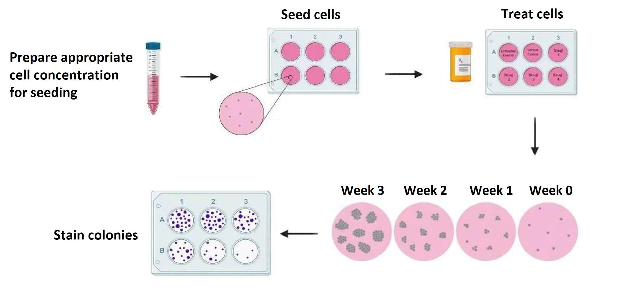 Fig.1.
Schematic representation of the clonogenic assay.
Fig.1.
Schematic representation of the clonogenic assay.
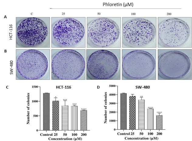 Fig.2.
Clonogenic assay. A, B HCT-116 and SW-480 cells treated with phloretin (25, 50, 100 200 µM), and stained with 0.5%
crystal violet. C, D Bar graphs showing relative colony growth of HCT-116 and SW-480 cells.[1]
Fig.2.
Clonogenic assay. A, B HCT-116 and SW-480 cells treated with phloretin (25, 50, 100 200 µM), and stained with 0.5%
crystal violet. C, D Bar graphs showing relative colony growth of HCT-116 and SW-480 cells.[1]
The soft agar colony formation assay is designed to assess the growth of anchorage-independent cells. This assay is particularly important for studying the transformation potential of cancer cells, as malignant cells can proliferate without the need for solid support.
It is essential for screening the tumorigenic potential of tumor cells and investigating the mechanisms of cancer cell growth and drug resistance, as it more closely mimics the environment of tumor growth compared to traditional two-dimensional cultures.
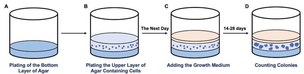 Fig.3.
Schematic representation of the soft agar colony formation assay.
Fig.3.
Schematic representation of the soft agar colony formation assay.
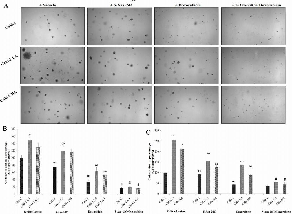 Fig.4.
Representative images (40X magnification) showing soft agar grown colonies as observed under microscope on day 14
after seeding (A), and histogram representation of numbers of colonies (B), and colony size (C), from Caki-1,
Caki-1LA and Caki-1HA cells treated with doxorubicin and 5-Aza-2dC either alone or their combination.[2]
Fig.4.
Representative images (40X magnification) showing soft agar grown colonies as observed under microscope on day 14
after seeding (A), and histogram representation of numbers of colonies (B), and colony size (C), from Caki-1,
Caki-1LA and Caki-1HA cells treated with doxorubicin and 5-Aza-2dC either alone or their combination.[2]
Service Features and Advantages
- Expertise and Experience: A team of skilled professionals with extensive experience in cellular assays, ensuring high-quality results.
- Assay Development and Optimization: Customized protocols designed to meet specific cell lines or experimental needs, with optimization for various conditions, including medium composition, growth factors, and assay duration.
- High Reproducibility: Rigorous quality control measures to guarantee consistent and reproducible assay outcomes.
- Speed and Efficiency: Streamlined processes that minimize turnaround time, facilitating timely results for experimental research.
- Cost-Effectiveness: Competitive pricing and a variety of service packages to accommodate different budget requirements.
FAQ
1. What is a colony formation assay?
A colony formation assay measures the ability of cells to grow into colonies. In this assay, a small number of cells are seeded onto a culture dish, and after a period of incubation, the resulting colonies are counted. This method is commonly used to study cell proliferation, survival, and the effects of drugs or treatments.
2: What cell types are suitable for this assay?
The assay works best with adherent cell lines but can also be adapted for suspension cells using appropriate methods, such as embedding them in a soft agar medium.
3: What are common challenges in performing the assay?
Uneven Seeding: Ensure that cells are evenly distributed during seeding to avoid variability.
Overlapping Colonies: Avoid high seeding densities that can lead to colony overlap.
Inconsistent Staining: Implement a consistent staining protocol to ensure visibility and reproducibility.
4: How can I optimize colony formation?
Use a low passage number of cells for consistency.
Optimize seeding density to prevent overcrowding or sparse plating.
Ensure proper incubation conditions, including temperature, humidity, and CO2 levels.
Validate the assay using controls, such as untreated versus treated groups.
5: How long does the assay take?
The assay duration depends on the doubling time of the cell line, typically 7-28 days, to allow sufficient colony growth.
Quotation and Ordering
If you have other questions, please contact us and we will provide you with detailed explanations. Our customer service representatives are available 24hr a day! Thanks for choosing Creative Bioarray services!
References
- Kapoor S, Padwad YS. Phloretin induces G2/M arrest and apoptosis by suppressing the β-catenin signaling pathway in colorectal carcinoma cells. Apoptosis. 2023;28(5-6):810-829. doi:10.1007/s10495-023-01826-4
- Ponnusamy L, Mahalingaiah PK, Singh KP. Chronic Oxidative Stress Increases Resistance to Doxorubicin-Induced Cytotoxicity in Renal Carcinoma Cells Potentially Through Epigenetic Mechanism. Mol Pharmacol. 2016;89(1):27-41. doi:10.1124/mol.115.100206
Explore Other Options