High-Fat Diet-Induced Nonalcoholic Fatty Liver Disease (NAFLD) Model
Creative Bioarray is a leading and reliable industry leader specializing in the meticulous development of animal models that accurately replicate human diseases. Among our extensive repertoire of models, we have successfully created the high-fat diet-induced nonalcoholic fatty liver disease (NAFLD) model. This model is designed to facilitate a deeper understanding of NAFLD's pathogenesis and to evaluate potential drug candidates.
The high-fat diet -induced NAFLD model is a pivotal tool in our preclinical research arsenal, designed to closely mimic the pathogenesis of human NAFLD. This model utilizes diets rich in fat to induce obesity and NAFLD in rodents. The model's efficacy in replicating metabolic syndrome, hepatic steatosis, and nonalcoholic steatohepatitis (NASH) has been well-validated, with variations in diet composition and duration enabling the study of different aspects of the disease.
Our High-fat Diet-Induced Nonalcoholic Fatty Liver Disease (NAFLD) Model
- Available Animal
- Rat
- Mouse
- Modeling Method
Animals are fed a high-fat diet for 16-20 weeks to induce NAFLD.
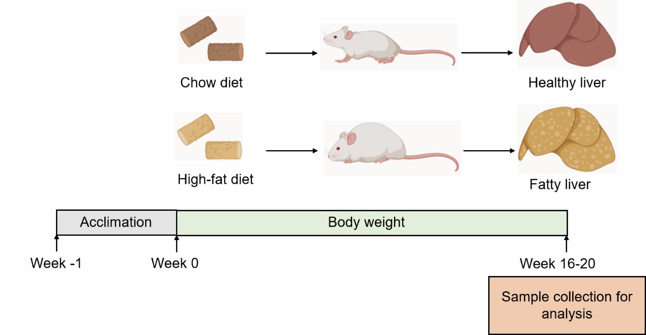 Fig. 1 Modeling method for the high-fat diet-induced NAFLD model at Creative Bioarray.
- Endpoints
- Body weight
- Liver weight
- Serum analysis: ALT, AST, TG, TC
- Histology analysis (liver): Oil red O staining
- Other customized endpoints
Example Data
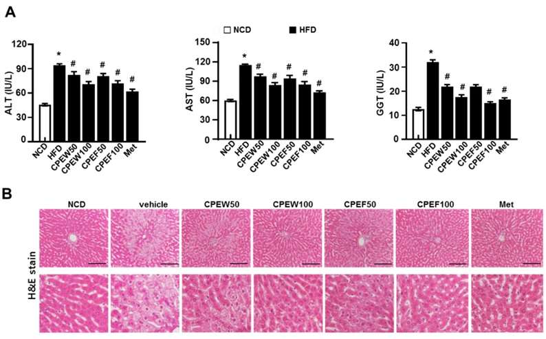 Fig. 2 Citrus peel extract protects against HFD-induced hepatic functional damage. (A) AST, ALT, and GGT were measured in HFD-fed rats. (B) H&E staining assay was performed using the liver. (Lee et al. 2020)
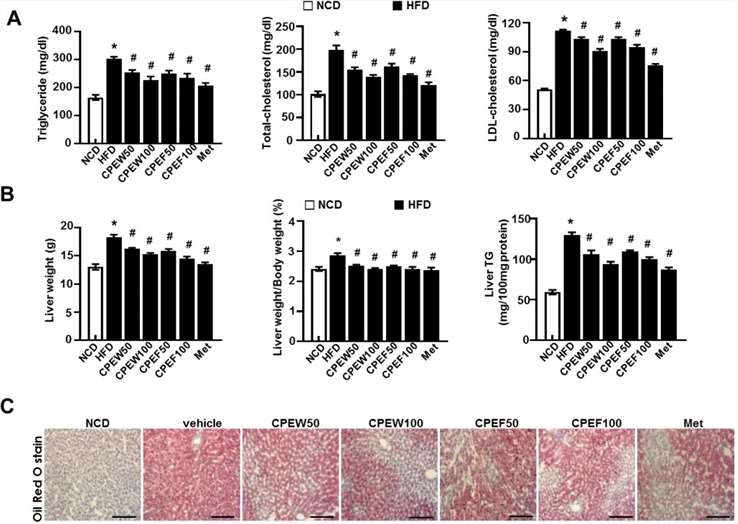 Fig 3 Citrus peel prevents HFD-induced hepatic steatosis. (A) Biochemical analysis of plasma samples. Levels of TG, TC and LDL-c were measured in the plasma of rat in different experimental groups. (B) Liver weight, and liver weight/body weight and liver TG content were measured 8 weeks after initial CPEW and CPEF administration. (C) Liver tissues were subjected to Oil Red O staining. (Lee et al. 2020)
Quotation and Ordering
Creative Bioarray is equipped with state-of-the-art preclinical technology platforms and backed by experienced research and scientific teams. Our comprehensive range of services is delivered with an unwavering commitment to high quality and efficiency, ensuring consistent exceptional high-quality results and rapid turnaround times. If you are interested in our services, please do not hesitate to contact us at any time or submit an inquiry to us directly.
References
- Fang, T., et al. Mouse models of nonalcoholic fatty liver disease (NAFLD): pathomechanisms and pharmacotherapies. International Journal of Biological Sciences, 2022, 18(15): 5681.
- Nevzorova, Y.A., et al. Animal models for liver disease–a practical approach for translational research. Journal of hepatology, 2020, 73(2): 423-440.
- Lee G H, Peng C, Park S A, et al. Citrus peel extract ameliorates high-fat diet-induced NAFLD via activation of AMPK signaling[J]. Nutrients, 2020, 12(3): 673.
For research use only. Not for any other purpose.
Disease Models
- Oncology Models
-
Inflammation & Autoimmune Disease Models
- Rheumatoid Arthritis Models
- Glomerulonephritis Models
- Multiple Sclerosis (MS) Models
- Ocular Inflammation Models
- Sjögren's Syndrome Model
- LPS-induced Acute Lung Injury Model
- Peritonitis Models
- Passive Cutaneous Anaphylaxis Model
- Delayed-Type Hypersensitivity (DTH) Models
- Inflammatory Bowel Disease Models
- Systemic Lupus Erythematosus Animal Models
- Oral Mucositis Model
- Asthma Model
- Sepsis Model
- Psoriasis Model
- Atopic Dermatitis (AD) Model
- Scleroderma Model
- Gouty Arthritis Model
- Carrageenan-Induced Air Pouch Synovitis Model
- Carrageenan-Induced Paw Edema Model
- Experimental Autoimmune Myasthenia Gravis (EAMG) Model
- Graft-versus-host Disease (GvHD) Models
-
Cardiovascular Disease Models
- Surgical Models
- Animal Models of Hypertension
- Venous Thrombosis Model
- Atherosclerosis model
- Cardiac Arrhythmia Model
- Hyperlipoidemia Model
- Doxorubicin-induced Heart Failure Model
- Isoproterenol-induced Heart Failure Model
- Arterial Thrombosis Model
- Pulmonary Arterial Hypertension (PAH) Models
- Heart Failure with Preserved Ejection Fraction (HFpEF) Model
- Cardio-Renal-Metabolic (CKM) Syndrome Model
-
Neurological Disease Models
- Alzheimer's Disease Modeling and Assays
- Ischemic Stroke Models
- Acute Spinal Cord Injury (ASCI) Model
- Traumatic Brain Injury (TBI) Model
- Hypoxic-Ischemic Encephalopathy (HIE) Model
- Tourette Syndrome (TS) Model
- Amyotrophic Lateral Sclerosis (ALS) Model
- Huntington's Disease (HD) Model
- Intracerebral hemorrhage (ICH) Models
- Schizophrenia Model
- Depression Models
- Pain Models
-
Metabolic Disease Models
- Type 1 Diabetes Mellitus Model
- Type 2 Diabetes Mellitus Model
- Animal Model of Hyperuricemia
-
Nonalcoholic Fatty Liver Disease Model
- High-Fat Diet-Induced Nonalcoholic Fatty Liver Disease (NAFLD) Model
- Methionine and Choline Deficient (MCD) Diet-Induced Nonalcoholic Fatty Liver Disease (NAFLD) Model
- Gubra-Amylin NASH (GAN) Diet-Induced Nonalcoholic Fatty Liver Disease (NAFLD) Model
- Streptozotocin (STZ) Induced Nonalcoholic Fatty Liver Disease (NAFLD) Model
- High Fat Diet-Induced Obesity Model
- Diabetic Foot Ulcer (DFU) Model
- Liver Disease Models
- Rare Disease Models
- Respiratory Disease Models
- Digestive Disease Models
-
Urology Disease Models
- Cisplatin-induced Nephrotoxicity Model
- Unilateral Ureteral Obstruction Model
- 5/6 Nephrectomy Model
- Renal Ischemia-Reperfusion Injury (RIRI) Model
- Diabetic Nephropathy (DN) Models
- Passive Heymann Nephritis (PHN) Model
- Adenine-Induced Chronic Kidney Disease (CKD) Model
- Kidney Stone Model
- Doxorubicin-Induced Nephropathy Model
- Orthotopic Kidney Transplantation Model
- Benign Prostatic Hyperplasia (BPH) Model
- Peritoneal Fibrosis Model
- Orthopedic Disease Models
- Ocular Disease Models
- Infectious Disease Models
- Skin Disease Models
- Otology Disease Models