Cell Angiogenesis Assays
- Service Details
- Features
- FAQ
Generate Predictive, Physiologically Relevant Data for Your Anti-Angiogenic Drug Discovery
In tumor progression and metastasis, angiogenesis is a critical, complex process often poorly modeled by conventional 2D systems. Move beyond limitations with our multi-dimensional angiogenesis platforms. We combine advanced in vitro 3D co-culture models with validated in vivo assays to mimic the tumor microenvironment, providing high-quality, reproducible data that translates more reliably to clinical outcomes. Accelerate your decision-making from early screening to preclinical validation.
Multi-Tiered Angiogenesis Platforms
In Vitro Platforms
HUVECs Tube Formation Assay
A rapid, quantitative assay to assess endothelial cell network formation on a basement membrane matrix.
- Physiologically Relevant Model: Utilizes primary human umbilical vein endothelial cells (HUVECs) on basement membrane matrix to closely recapitulate in vivo angiogenic processes, delivering biologically meaningful results that translate well to preclinical and clinical stages.
- Your Benefit: Obtain high-throughput, sensitive data on compound effects within days, ideal for primary screening.
- Screening Formats: 96-/384-well plates; High-content imaging; Multi-endpoint options (tube formation + viability + apoptosis).
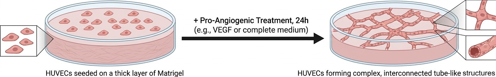 Fig. 1. Tube formation assay.
Case Study
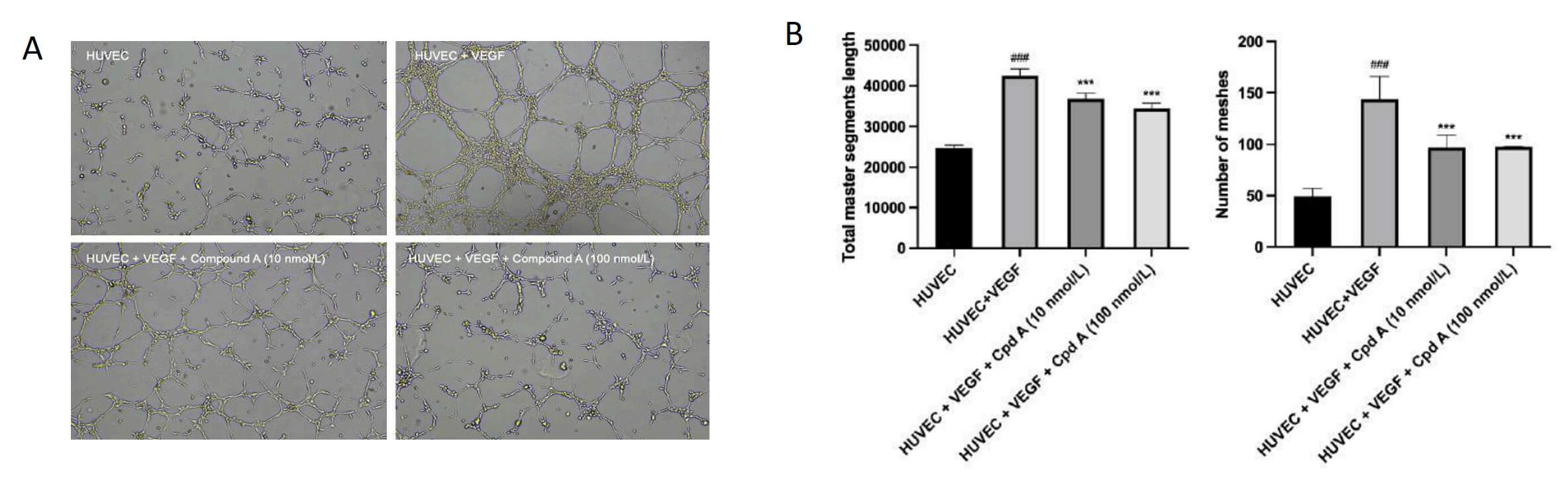 Fig. 2. (A) The effects of test compound A on VEGF-induced HUVECs tube formation. (B) Quantification of cell angiogenesis by counting the total master segments length and the number of meshes.
Spheroid Sprouting Assay
A 3D model where endothelial spheroids embedded in collagen/Matrigel form sprouts, measuring angiogenic invasion.
- Enhanced Physiological Model: Co-culture with tumor spheroids reproduces critical microenvironmental cues and paracrine signaling.
- Your Benefit: Capture complex 3D interactions and sprouting dynamics predictive of in vivo vascular remodeling for more confident candidate selection.
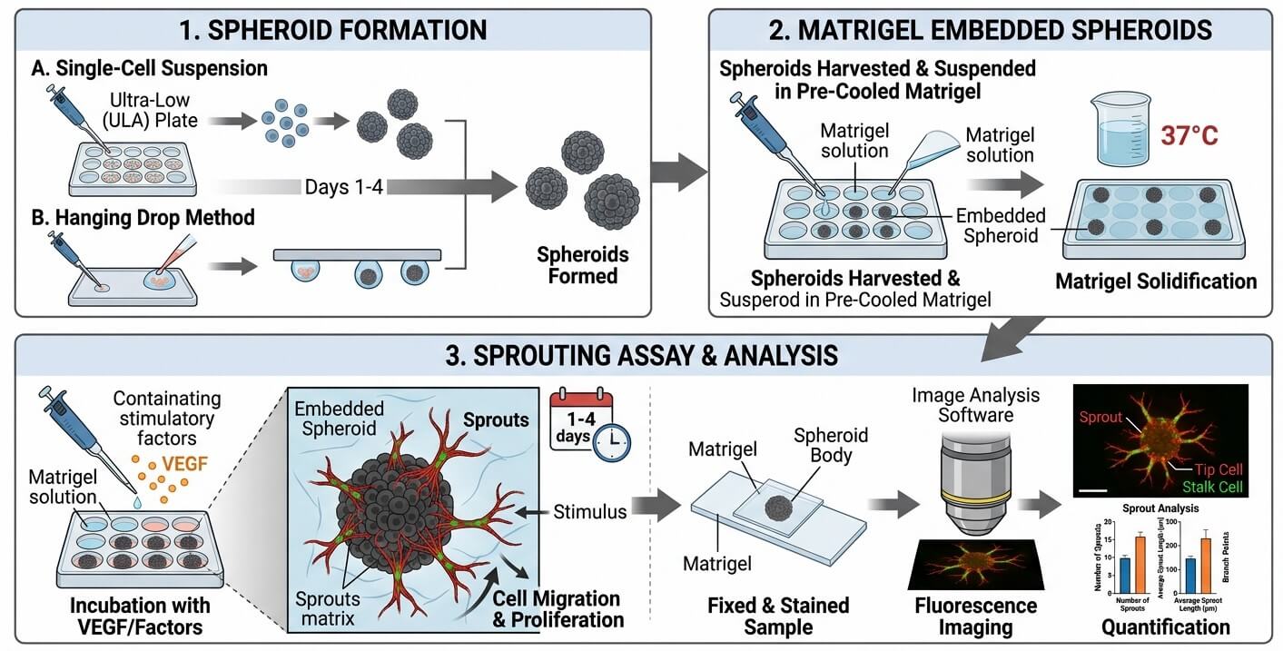 Fig. 3. Spheroid sprouting assay.
Rat Aortic Ring Assay
An ex vivo model monitoring microvessel outgrowth from primary aortic tissue, offering high physiological relevance.
- Enhanced Physiological Model: Can be stimulated with tumor-conditioned media to simulate tumor-induced angiogenesis.
- Your Benefit: Gain mechanistic insight into angiogenic sprouting and compound effects on primary tissue with intact cellular heterogeneity and extracellular matrix.
- Screening Formats: 24-/48-well plates; Real-time, live-cell imaging.
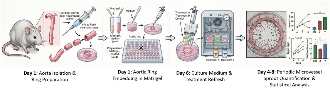 Fig. 4. The workflow of rat aortic ring assay.
While angiogenesis assays evaluate vascular formation, tumor progression also involves endothelial migration and extracellular matrix invasion. To provide a more comprehensive functional profile of your compounds, we also offer:
Quantitative assessment of tumor or endothelial invasion within 3D matrices.
Evaluation of chemotactic migration under controlled gradient conditions.
Ex Vivo & In Vivo Platforms
Matrigel Plug Assay
A standard, quantitative in vivo assay where subcutaneous Matrigel plugs are used to assess functional angiogenesis.
- Enhanced Physiological Model: Plugs can be formulated with tumor cells or tumor-conditioned media to model tumor angiogenesis.
- Your Benefit: Confirms compound efficacy in a live animal model with robust, quantitative endpoints, bridging in vitro and in vivo findings.
- Screening Formats: Individual plugs per condition; Multi-parameter quantitative analysis.
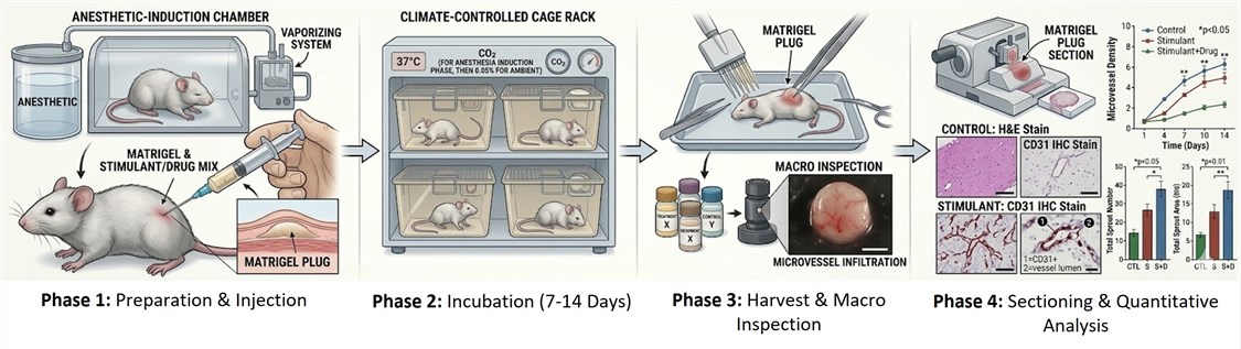 Fig. 5. The workflow of Matrigel plug assay.
CAM (Chick Chorioallantoic Membrane) Assay
A rapid, cost-effective in vivo assay using the vascular chick embryo membrane for high-throughput screening.
- Enhanced Physiological Model: Tumor tissue grafts can be placed on the CAM to study angiogenesis in a tumor microenvironment.
- Your Benefit: Rapidly screen multiple compounds in vivo with clear visualization of vascular effects, optimizing resource allocation.
- Screening Formats: Multi-embryo screening; Semi-automated image analysis.
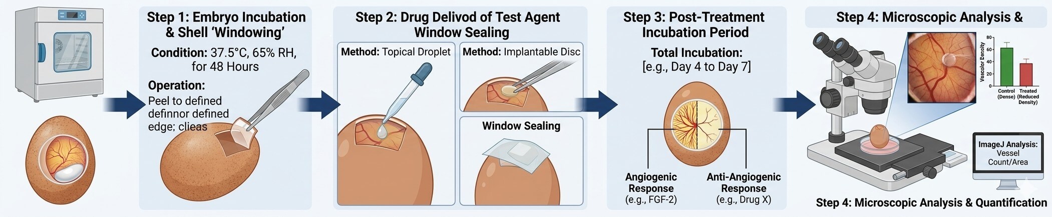 Fig 6. Optimized chick chorioallantoic membrane (CAM) angiogenesis assay.
Case Study
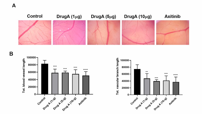 Fig 7. Activity of drug A in CAM angiogenesis assay. (A) The representative microscopic images of CAM angiogenesis after 24 h of incubation with different treatments. (B) Quantification of CAM angiogenesis by measuring the total length of the blood vessels and blood vessel branches.
Corneal Angiogenesis Assay
A highly sensitive in vivo model inducing and quantifying neovascularization in the normally avascular cornea.
- Your Benefit: Provides a precise, localized, and quantitative measure of angiogenic response, ideal for validating potent modulators in a sensitive site.
- Screening Formats: Single compound per eye; Detailed morphometric analysis.
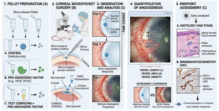 Fig. 8. Corneal angiogenesis Assay
Our Delivery & Support
To achieve the best outcomes, we recommend providing:
- Compound information: type, solubility, stability, and intended dosing range.
- Optional cell lines or samples: customer-supplied cells can be accommodated with tailored protocols.
- Desired endpoints: viability, tube formation, sprouting, or migration metrics.
You will receive:
- Comprehensive PDF report with methodology, representative images, and quantitative analysis
- All raw images and data files for independent review
- Multi-endpoint analysis and statistical figures for actionable insights
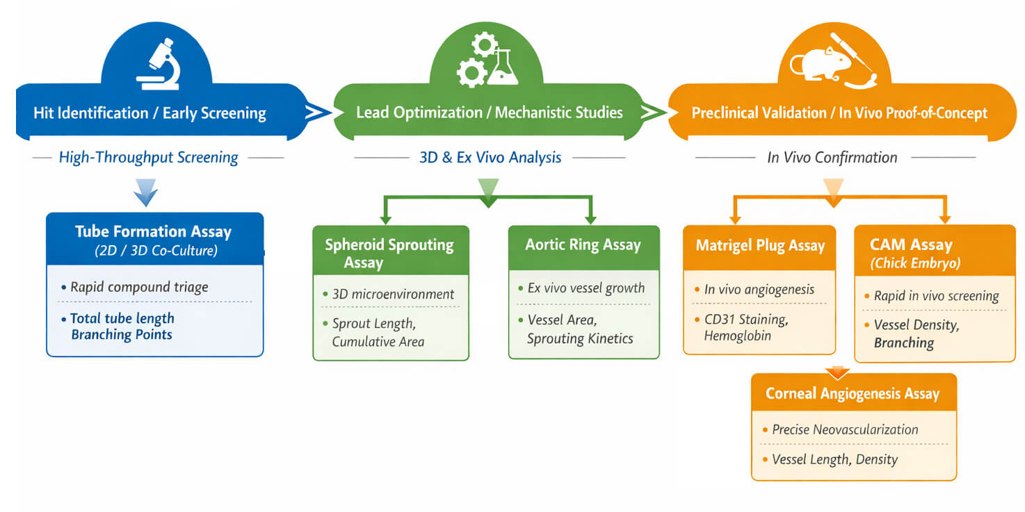
Not Sure Which Assay to Choose?
We recommend selecting angiogenesis assays based on the stage of your drug discovery program.
Why Choose Creative Bioarray

Advanced & Physiologically Relevant Models
We integrate 2D, 3D, ex vivo, and in vivo platforms, including 3D co-culture and tumor microenvironment–mimicking systems, to deliver more predictive angiogenesis data.

Reliable and Reproducible Data
All assays follow standardized SOPs with rigorous controls and quantitative analysis, ensuring consistent and decision-ready results.

Flexible for Complex Compounds
Customized protocols support diverse study designs and challenging compounds, including poorly soluble or volatile molecules.

End-to-End Decision Support
From high-throughput screening to in vivo validation, our platforms support every stage of oncology drug discovery.
FAQ
What type of samples or information should I send?
Please provide information on the compounds you would like to test (compound type, solubility, stability, desired dosing). If you are planning to use customer-supplied cells, please send those as well. Let us know what endpoints you are most interested in so we can design the assay to meet your needs.
Is it possible to modify assays to meet special requirements?
Absolutely. Cell types, dosing schedules, inclusion of multi-endpoint readouts, and specialized handling of compounds can all be tailored to meet the needs of your project.
What do I receive?
You receive a PDF report complete with original images, data files and statistical analysis. Data is delivered ready to share with outside collaborators, use in presentations or publish.
I have some compounds that are not very soluble. How can this be addressed?
Our formulation platform allows us to choose the best solvent or media to get your compound adequately dispersed in our 3D matrices.
Call to Action
Ready to accelerate your early drug discovery? Contact us today to discuss your project and get a tailored angiogenesis assay plan that delivers actionable, reliable, and high-quality data.
Explore Other Options