Liver Fibrosis Model
Liver fibrosis is a wound healing response to insults and is the final common pathway of chronic liver disease of various etiologies, which can progress into more severe stages, such as liver cirrhosis, liver failure and hepatocellular carcinoma. The activation of hepatic stellate cells, portal fibroblasts, and myofibroblasts of bone marrow origin is the most important event in liver fibrosis. Currently, lots of patients suffer from liver fibrosis because of alcohol abuse, chronic hepatitis virus infection and non-alcoholic steatohepatitis worldwide.
Animals of liver fibrosis are used as models to study liver fibrosis in human and test subjects for the development and testing of drugs, vaccines, and other biologicals.
Creative Bioarray specializes in providing customized pharmacodynamic research services to help customers assess the efficacy of drug candidates and study the associated pathological mechanisms through liver fibrosis models.
Animal models of liver fibrosis include but not limited to the following:
- Chemical-Induced Liver Fibrosis Models
- Carbon Tetrachloride-Induced Liver Fibrosis Model
- Thioacetamide-Induced Liver Fibrosis Model
- Diet-Induced Liver Fibrosis Models
- Methionine-Deficient and Choline-Deficient Diet-Induced Liver Fibrosis Model
- Genetically Modified Models
- Multidrug Resistance-Associated Protein 2-Deficient Mice
- Alms1Fat Ausi Mutant Mice
Species available
- Rat
- Mouse
Our capabilities
- Liver fibrosis in animal is induced by twice weekly intraperitoneal injections of CCl4.
- We evaluate the degree of liver fibrosis by Hematoxylin and Eosin (H&E) and Masson's trichrome.
- We assess the apoptosis of the paraffin-embedded liver sections of mice/rat by TUNEL staining and immunofluorescent staining.
Assays available
- Biochemical analysis
- Pathological evaluation
With extensive experience in the field of liver fibrosis, we are confident to help you overcome any upcoming challenges. Our experts are fully capable of customizing our protocols and assays to meet your specific needs. With our help, we wish to facilitate your research with high efficiency.
Study examples
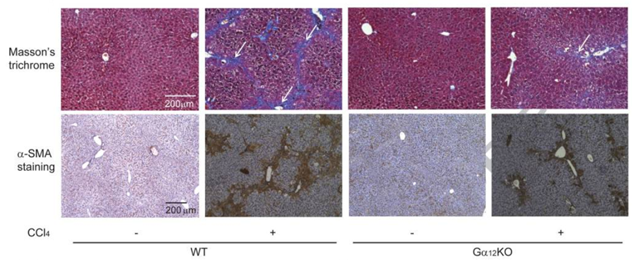 Figure. 1. Liver fibrosis is inhibited by Gα12KO. Masson’s trichrome staining and α-SMA staining of liver tissue of different treatment group.
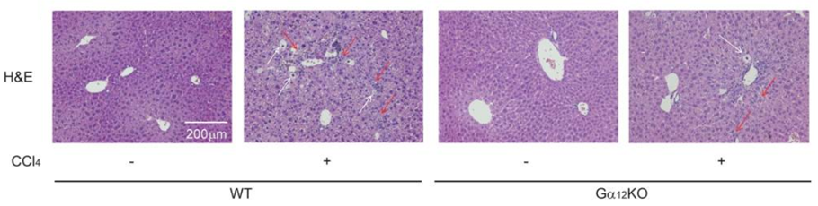 Figure. 2. Liver fibrosis is inhibited by Gα12KO. H&E staining of liver tissue of different treatment group.
Quotation and ordering
If you have any special needs or questions regarding our services, please feel free to contact us. We look forward to cooperating with you in the future.
Reference
Kyu Min Kim, et al. Gα12 overexpression induced by miR-16 dysregulation contributes to liver fibrosis by promoting autophagy in hepatic stellate cells[J]. Journal of Hepatology: The Journal of the European Association for the Study of the Liver, 2018.
For research use only. Not for any other purpose.
Disease Models
- Oncology Models
-
Inflammation & Autoimmune Disease Models
- Rheumatoid Arthritis Models
- Glomerulonephritis Models
- Multiple Sclerosis (MS) Models
- Ocular Inflammation Models
- Sjögren's Syndrome Model
- LPS-induced Acute Lung Injury Model
- Peritonitis Models
- Passive Cutaneous Anaphylaxis Model
- Delayed-Type Hypersensitivity (DTH) Models
- Inflammatory Bowel Disease Models
- Systemic Lupus Erythematosus Animal Models
- Oral Mucositis Model
- Asthma Model
- Sepsis Model
- Psoriasis Model
- Atopic Dermatitis (AD) Model
- Scleroderma Model
- Gouty Arthritis Model
- Carrageenan-Induced Air Pouch Synovitis Model
- Carrageenan-Induced Paw Edema Model
- Experimental Autoimmune Myasthenia Gravis (EAMG) Model
- Graft-versus-host Disease (GvHD) Models
-
Cardiovascular Disease Models
- Surgical Models
- Animal Models of Hypertension
- Venous Thrombosis Model
- Atherosclerosis model
- Cardiac Arrhythmia Model
- Hyperlipoidemia Model
- Doxorubicin-induced Heart Failure Model
- Isoproterenol-induced Heart Failure Model
- Arterial Thrombosis Model
- Pulmonary Arterial Hypertension (PAH) Models
- Heart Failure with Preserved Ejection Fraction (HFpEF) Model
- Cardio-Renal-Metabolic (CKM) Syndrome Model
-
Neurological Disease Models
- Alzheimer's Disease Modeling and Assays
- Seizure Models
- Parkinson's Disease Models
- Ischemic Stroke Models
- Acute Spinal Cord Injury (ASCI) Model
- Traumatic Brain Injury (TBI) Model
- Hypoxic-Ischemic Encephalopathy (HIE) Model
- Tourette Syndrome (TS) Model
- Amyotrophic Lateral Sclerosis (ALS) Model
- Huntington's Disease (HD) Model
- Intracerebral hemorrhage (ICH) Models
- Schizophrenia Model
- Depression Models
- Pain Models
-
Metabolic Disease Models
- Type 1 Diabetes Mellitus Model
- Type 2 Diabetes Mellitus Model
- Animal Model of Hyperuricemia
-
Nonalcoholic Fatty Liver Disease Model
- High-Fat Diet-Induced Nonalcoholic Fatty Liver Disease (NAFLD) Model
- Methionine and Choline Deficient (MCD) Diet-Induced Nonalcoholic Fatty Liver Disease (NAFLD) Model
- Gubra-Amylin NASH (GAN) Diet-Induced Nonalcoholic Fatty Liver Disease (NAFLD) Model
- Streptozotocin (STZ) Induced Nonalcoholic Fatty Liver Disease (NAFLD) Model
- High Fat Diet-Induced Obesity Model
- Diabetic Foot Ulcer (DFU) Model
- Cardio-Renal-Metabolic (CKM) Syndrome Model
- Liver Disease Models
- Rare Disease Models
- Respiratory Disease Models
- Digestive Disease Models
-
Urology Disease Models
- Cisplatin-induced Nephrotoxicity Model
- Unilateral Ureteral Obstruction Model
- 5/6 Nephrectomy Model
- Renal Ischemia-Reperfusion Injury (RIRI) Model
- Diabetic Nephropathy (DN) Models
- Passive Heymann Nephritis (PHN) Model
- Adenine-Induced Chronic Kidney Disease (CKD) Model
- Kidney Stone Model
- Doxorubicin-Induced Nephropathy Model
- Orthotopic Kidney Transplantation Model
- Benign Prostatic Hyperplasia (BPH) Model
- Peritoneal Fibrosis Model
- Cardio-Renal-Metabolic (CKM) Syndrome Model
- Orthopedic Disease Models
- Ocular Disease Models
- Infectious Disease Models
- Skin Disease Models
- Otology Disease Models