Comprehensive Comparison of IHC, CISH, and FISH Techniques
Immunohistochemistry (IHC), chromogenic in situ hybridization (CISH), and fluorescence in situ hybridization (FISH) are pivotal techniques in life sciences. Each method offers unique advantages and applications for visualizing specific biomolecules within cells and tissues.
Comparison of Principles and Mechanisms
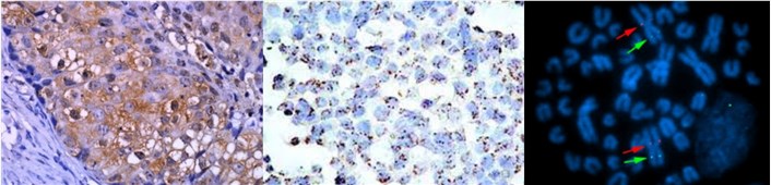
- IHC. It involves the use of antibodies conjugated to an enzyme or a fluorophore to detect and visualize specific proteins within cells and tissues. This technique relies on the binding of antigen-specific antibodies to target proteins, followed by the detection of antibody-antigen complexes using enzyme-mediated chromogenic reactions or direct fluorescence from fluorophore-conjugated antibodies. The visualization of these complexes provides valuable insights into protein expression, cellular localization, and tissue morphology, forming the basis for diverse applications in research and diagnostics.
- CISH. It is a specialized technique that utilizes labeled nucleic acid probes to visualize specific DNA or RNA sequences within cells and tissues. Its detection method involves the enzymatic conversion of a chromogenic substrate to produce a visible signal at the site of hybridization, allowing for the precise localization of nucleic acid targets within the cellular context. CISH has emerged as a valuable tool in cancer diagnostics and research, enabling the detection of gene amplifications, translocations, and other genetic aberrations with high specificity and sensitivity.
- FISH. It employs fluorescently labeled nucleic acid probes to visualize specific DNA or RNA sequences within fixed cells and tissues. This technique harnesses the visualization of fluorescent signals under a fluorescence microscope, facilitating the precise localization and quantification of nucleic acid targets. Its versatility allows for the simultaneous visualization of multiple targets through multicolor FISH, making it indispensable in genetics, cytogenetics, oncology, and microbiology research.
Comparison of Preparation and Analysis
| Techniques | Preparation work | Analysis work |
| IHC | Tissue fixation, embedding, sectioning, deparaffinization, antigen retrieval, blocking, antibody incubation, detection, counterstaining, and mounting. | After the completion of the staining and mounting process, the tissue sections are examined under a microscope. The specific antigenic sites that have interacted with the antibodies will display a visible color change or fluorescence. The pattern and distribution of staining are analyzed to determine the spatial localization of the target antigen within the tissue sample. The intensity of staining is evaluated, typically using a semi-quantitative scale, to assess the relative abundance or expression level of the target antigen. |
| CISH | Tissue fixation, embedding, and sectioning, deparaffinization, pre-hybridization, hybridization, stringency washes, detection. | The tissue sections are observed under a bright-field microscope to detect the presence of chromogenic signals that indicate the location of the target DNA or RNA sequences. The number of signals per cell, their distribution, and any specific patterns are assessed to characterize the genetic aberrations or gene amplifications present in the tissue sample. |
| FISH | Tissue fixation, embedding, and sectioning, pre-hybridization, hybridization, stringency washes, staining, mounting. | Using a fluorescence microscope, the tissue sections are visualized to detect the fluorescent signals corresponding to the hybridized probes. Digital image analysis systems may be employed to quantify the signals and provide more objective measurements, such as the ratio of different fluorescent signals. |
Sensitivity and Specificity
- IHC. It has moderate to high sensitivity in detecting specific proteins, depending on the quality of antibodies used and the target protein's abundance within the sample. This approach provides high specificity in detecting and localizing specific protein targets, as it relies on the specificity of antigen-antibody interactions.
- CISH. It offers high sensitivity in visualizing specific DNA or RNA sequences within cells and tissues, providing valuable insights into genetic aberrations and alterations. This method offers high specificity in visualizing specific nucleic acid sequences, enabling precise localization of genetic alterations within the cellular context.
- FISH. It is known for its exceptional sensitivity, surpassing the sensitivity of IHC and CISH due to the bright fluorescence signals and the absence of background noise associated with enzyme-mediated reactions. This technique demonstrates superior specificity in localizing specific DNA or RNA sequences within cells and tissues, enhancing the accuracy of its analytical outputs in diverse research and diagnostic applications.
Multiplexing Capabilities
- IHC. It does support multiplexing, allowing the simultaneous detection and visualization of multiple proteins within a single sample. However, it is limited by spectral overlap in the case of fluorescent detection and substrate constraints in chromogenic detection, which may affect the number of targets that can be simultaneously visualized.
- CISH. It supports multiplexing, enabling the visualization of multiple DNA or RNA targets within cells and tissues. Similar to IHC, it is constrained by the spectral overlap in fluorescent detection and substrate limitations in chromogenic detection.
- FISH. It excels in multiplexing capabilities, allowing the visualization of multiple nucleic acid targets within the same sample. It offers the unique capacity for multicolor FISH, enabling the simultaneous detection and visualization of different DNA or RNA sequences with distinct fluorescent labels. This capability provides researchers with a comprehensive view of chromosomal rearrangements, gene expression networks, and spatial relationships between genomic loci.
Creative Bioarray Relevant Recommendations
| Product/Service Types | Description |
| Immunohistochemistry (IHC), Immunofluorescence (IF) Service | Creative Bioarray offers a comprehensive IHC service from project design, and marker selection to image completion and data analysis. We are dedicated to satisfying every customer and assisting them to achieve their specific research goals. |
| Fluorescent In Situ Hybridization (FISH) Service | Creative Bioarray offers a range of different FISH services including metaphase and interphase FISH, fibre-FISH, RNA-FISH, M-FISH, 3D-FISH, flow-FISH, FISH on paraffin sections, and immune-FISH. |
| FISH Probe Design, Synthesis, and Testing Service | Creative Bioarray is capable of developing custom FISH probes. Apart from that, we also offer mRNA ISH/FISH probes, miRNA ISH/FISH probes, and lncRNA ISH/FISH probes. |
| IHC ISH Reagents | Creative Bioarray specializes in the provision of a diverse range of high-quality IHC and ISH reagents. |