Small RNA Detection by ISH Methods
Small RNAs, including microRNAs (miRNAs) and small interfering RNAs (siRNAs), are critical players in gene regulation and have emerged as key molecules in various biological processes. Detecting and characterizing small RNAs is crucial for understanding their functional roles and their implications in health and disease. In situ hybridization (ISH) methods provide powerful tools for visualizing and studying small RNA molecules within cells and tissues.
Principles of ISH for Small RNA Detection
ISH for small RNA detection relies on the hybridization of small RNA probes with complementary target sequences. These probes are designed to specifically bind to the small RNA molecules of interest. The probes are labeled with fluorescent dyes or enzyme-based labels, enabling their visualization. Signal amplification strategies can be employed to enhance sensitivity, ensuring the detection of even low-abundance small RNA molecules.
The hybridization process involves the denaturation of target RNA molecules, allowing the probes to hybridize and form stable complexes. The specificity of the hybridization reaction is achieved through stringent washing steps, removing non-specifically bound probes. The remaining probes bound to their complementary targets generate signals that can be visualized and quantified.
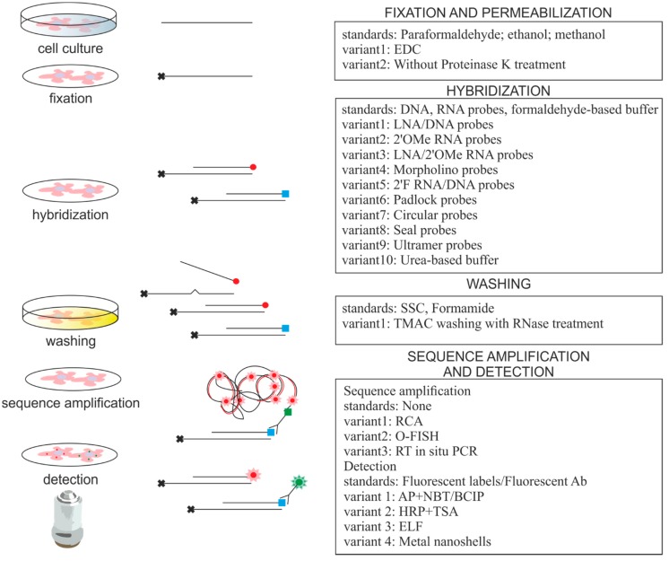 Fig. 1 In situ hybridization protocols used for imaging of small RNAs. (Urbanek MO, et al., 2015)
Design and Synthesis of Small RNA Probes
Designing effective small RNA probes is crucial for accurate and specific detection. Several considerations come into play during probe design. Probes are typically designed to target regions of the small RNA molecule that exhibit high conservation across species or are unique to the target small RNA.
Factors such as probe length, sequence selection, and secondary structure influence the efficiency and specificity of hybridization. Longer probes may provide increased sensitivity but can also introduce non-specific binding. Careful selection of probe sequences, avoiding regions prone to secondary structure formation, helps enhance the specificity of the hybridization reaction.
Probes can be labeled with fluorescent dyes, such as cyanine dyes or Alexa Fluor dyes, enabling visualization using fluorescence microscopy. Alternatively, enzyme-based labels, such as horseradish peroxidase (HRP) or alkaline phosphatase (AP), can be employed in chromogenic ISH (CISH) techniques, leading to colorimetric visualization.
Variations in Small RNA ISH Techniques
Small RNA ISH techniques encompass various methods tailored to specific experimental requirements. Two commonly used techniques are fluorescent ISH (FISH) and chromogenic ISH (CISH).
- Fluorescent ISH (FISH) for small RNA detection
FISH allows for the visualization of small RNA molecules in cells and tissues using fluorescently labeled probes. Fluorescent dyes, such as Cy3, Cy5, or fluorescein, are commonly used to label the probes, emitting distinct colors when excited by specific wavelengths of light. This technique enables the simultaneous detection of multiple small RNA targets, facilitating the investigation of complex regulatory networks.
FISH can provide valuable insights into the cellular localization and abundance of small RNAs. It allows researchers to examine the spatial distribution patterns of small RNAs within tissues, identifying specific cell types or subcellular compartments where they are enriched. Furthermore, FISH can be combined with immunohistochemistry (ISH-IHC) to co-detect small RNAs and proteins, offering a comprehensive understanding of their interplay in cellular processes. - Chromogenic ISH (CISH) for small RNA visualization
CISH techniques employ enzyme-based labels to visualize small RNA molecules. The hybridization of probes to target small RNAs is detected through the enzymatic conversion of chromogenic substrates, resulting in the formation of colored precipitates. Commonly used enzyme labels include horseradish peroxidase (HRP) and alkaline phosphatase (AP).
CISH provides permanent staining, allowing the preservation of the signal for long-term observation. It offers excellent spatial resolution and compatibility with standard histological techniques. CISH is particularly useful when archival tissue sections or large-scale tissue arrays are employed, making it a practical approach for retrospective studies and clinical applications.
Creative Bioarray Relevant Recommendations
Creative Bioarray is a leading biological company with extensive expertise in the field of microRNA research. With a strong emphasis on innovation, we specialize in the development and production of high-quality microRNA probes.
| Cat. No. | Product Name |
| FMRC-01 | miRprobe Detection Probe |
| FMRC-02 | microRNA-122a Probe |
| FMRC-03 | microRNA-124a Probe |
| FMRC-04 | microRNA-138 Probe |
| FMRC-05 | microRNA-206 Probe |
| FMRC-06 | microRNA-375 Probe |
| FMRC-07 | microRNA-377 Probe |
| FMRC-08 | microRNA-93 Probe |
View the details of our microRNA probes and find what you need!
Reference
- Urbanek MO, et al. (2015). "Small RNA Detection by in Situ Hybridization Methods." Int J Mol Sci. 16 (6), 13259-86.