How Immunohistochemistry Makes the Invisible Brain Visible?
Immunohistochemistry (IHC) allows for the localisation, qualitative and semi-quantitative analysis of particular proteins or molecules of interest within tissue or cells based on antigen–antibody specificity. The high specificity and spatial resolution of IHC have made it one of the fundamental techniques in neuroscience for understanding neuronal structure, function, and mechanisms of disease. Broadly, its four most common applications in neuroscience are: identifying neurons and subcellular specialisations; mapping neural circuits; studying the regulation of functional proteins; and investigating neuropathological mechanisms.

Identification of Neural Cell Types and Subcellular Structures
The nervous system contains many different cell types including neurons, glial cells (astrocytes, oligodendrocytes, microglia) and vascular endothelial cells that can be identified based on their expression of characteristic molecular markers. IHC allows these markers to be recognised and used to precisely identify cell types or localise subcellular structures.
Common Markers for Neural Cell Identification
| Cell Type | Representative Markers (Antibodies) | Description |
|---|---|---|
| Neurons (mature) | NeuN (RBFOX3), MAP2, βIII-Tubulin (TUBB3), NSE, Neurofilament (NF), Synaptophysin (Syn) | NeuN labels neuronal nuclei and is widely used for neuron counting. MAP2 and βIII-Tubulin label dendrites and axons, respectively. NSE, NF, and Syn serve as complementary functional/structural markers. |
| Astrocytes | GFAP, S100β, ALDH1L1 (optional) | GFAP is the classical cytoskeletal marker for astrocytes; S100β is often co-stained with GFAP to increase specificity. |
| Oligodendrocytes / Myelinating Cells | Olig2, MBP, MOG, MAG | Olig2 labels oligodendrocyte lineage cells; MBP, MOG, and MAG mark myelin sheaths directly. |
| Microglia / Brain-Resident Macrophages | Iba1, TMEM119, CD68 (optional) | Iba1 and TMEM119 are specific for microglia, with TMEM119 showing higher specificity in mature cells. |
| Neural Stem / Progenitor Cells | Nestin, SOX2, CD133 (optional) | Nestin and SOX2 identify undifferentiated neural stem or progenitor cells. |
Subcellular Structural Markers
| Structure | Representative Markers | Description |
|---|---|---|
| Axon | Neurofilament (NF-L/M/H), βIII-Tubulin, Tau, Ankyrin-G (axon initial segment) | NF proteins indicate axonal integrity; βIII-Tubulin and Tau are used to assess axonal growth or degeneration. |
| Dendrite | MAP2, PSD-95, Shank, Homer1 | MAP2 labels dendritic microtubules; PSD-95 and Shank are postsynaptic density markers suitable for studying synaptic plasticity. |
| Presynaptic Terminal | Synaptophysin, Synapsin I, Bassoon, Piccolo | Synaptophysin and Synapsin I mark synaptic vesicles; Bassoon and Piccolo define the presynaptic active zone. |
| Postsynaptic Region | PSD-95, Homer1, Gephyrin (inhibitory synapses) | These proteins help distinguish excitatory versus inhibitory synaptic sites. |
| Nucleus / Proliferation | Ki-67, PCNA, p53 | Ki-67 and PCNA indicate proliferative activity, especially in neural stem cells or tumor models. |
| Mitochondria | COX IV, TOM20 | Used to study energy metabolism and cell death pathways. |
| Endoplasmic Reticulum | Calnexin, PDI | Indicators of ER stress and protein folding capacity. |
| Lysosome | LAMP1 | Used to assess autophagic or lysosomal activity. |
Neural Circuit Construction and Mapping
Neural circuits are the structural basis of various brain functions, including perception, movement, and memory. IHC (often combined with double or multiple labeling and FISH) can be applied to label neurotransmitter/receptor markers and neuronal markers to reveal interregional connectivity and the type of neurotransmission.
Localization of Projection Neurons Across Brain Regions
For example, fluorescent retrograde tracers (e.g., Fluoro-Gold) can be injected into the cortex for retrograde tracing of the cortex–hippocampus memory circuit, and then IHC staining for NeuN (neurons) and VGLUT1 (excitatory neurons). Dual labeling would reveal not only the identity of the projecting neurons, but also the neurotransmitter type, and precisely map their spatial distribution across the circuit.
Synaptic Connection Typing
Dual labeling with presynaptic (Synaptophysin) and postsynaptic markers (PSD-95) allows for direct visualization of excitatory synaptic morphology and density under the microscope, revealing structural plasticity associated with learning and memory.
Expression and Regulation of Molecules Related to Neural Function
Neural functions such as neurogenesis, synaptic plasticity, and stress response depend on dynamic spatiotemporal protein expression. Semi-quantitative IHC analyses (average optical density or percentage of positive cells) can uncover the changes in expression under physiological or pathological conditions and help to relate molecular regulation to neural function.
- Neurogenesis Studies: Dual IHC for BrdU (proliferating cells) and NeuN (mature neurons) in the adult hippocampal dentate gyrus can be used to quantify newborn neurons and assess how drugs (e.g., antidepressants) or environmental enrichment regulate neurogenesis.
- Neurotransmitter Receptor Distribution: Detection of NMDA receptor subunit GluN1 in the hippocampus can reveal changes in distribution associated with long-term potentiation (LTP), a cellular process that underlies memory formation.
- Stress-Response Molecules: IHC analysis of CRH and GR expression along the hypothalamic–pituitary–adrenal (HPA) axis can be used to help understand how stress affects central nervous system homeostasis.
Pathological Mechanisms in Neurodegenerative Diseases and Brain Injury
Neurodegenerative Diseases
| Pathological Process | Key Markers (Common Antibodies) | Research Insight / Typical Findings |
|---|---|---|
| Protein Misfolding and Aggregation | Aβ, phospho-Tau, α-Synuclein, mutant Huntingtin (mHtt), TDP-43 | Quantifying plaques and tangles by IHC assists in staging Alzheimer's and Parkinson's disease; plaque density correlates positively with cognitive impairment. |
| Neuroinflammation and Immune Cells | Iba1, TMEM119 (microglia), GFAP, S100β (astrocytes), CD68, CD3, Granzyme K⁺ CD8 T cells | Iba1/TMEM119 staining highlights microglial activation; CD8 T cell infiltration has been linked to modulating tau pathology, indicating a dual role of adaptive immunity. |
| Cell Death and Stress Pathways | Cleaved-caspase-3, PARP-1, 4-HNE, 8-OH-dG, LC3-II, p62 | IHC demonstrates neuronal apoptosis and oxidative stress aggregation in AD and ALS, helping map temporal and spatial cell-death patterns. |
| Immune-Mediated Secondary Damage | Eomes⁺ Th cells, LINE-1 ORF1 protein | Eomes⁺ Th cells recognize aberrant LINE-1 ORF1 expression in neurons, initiating immune-dependent neuronal injury — a two-stage "intrinsic death plus immune damage" model. |
| Disease-Specific Signaling Pathways | TLR4, NF-κB, p-STAT3 | Upregulation of TLR4/NF-κB signaling in microglia demonstrates that innate immune activation is a shared pathogenic driver in AD and PD. |
Brain Injury
| Injury Phase | Key Markers | Representative Findings |
|---|---|---|
| Primary Mechanical Injury | βIII-Tubulin, NeuN | In traumatic brain injury (TBI) models, rapid loss of NeuN-positive cells indicates acute neuronal death. |
| Secondary Inflammation | Iba1, TMEM119, GFAP, AQP4, TNF-α, IL-1β | In MCAO (ischemia) models, increased AQP4 expression marks cerebral edema; localized TNF-α/IL-1β expression identifies inflammatory hotspots. |
| Blood–Brain Barrier Breakdown | ZO-1, Claudin-5, Fibrinogen | Fibrinogen deposition in brain parenchyma reflects BBB leakage after hemorrhage, quantifiable via IHC. |
| Oxidative Stress and Ferroptosis | 4-HNE, MDA, GPX4, COX-2, TfR, Fpn1 | Post-hemorrhagic iron accumulation increases ROS and reduces GPX4, with IHC mapping ferroptosis markers across time and space. |
| Immune Cell Infiltration | CD8⁺ T cells, Granzyme K⁺ T cells | In febrile seizure models, hippocampal TNF-α and IL-1β expression correlates strongly with CD8⁺ T cell infiltration, implicating adaptive immunity in secondary damage. |
| Cell Adhesion and Apoptosis | Lnx1, caspase-3, Bax/Bcl-2 | In TBI cortex, Lnx1 upregulation contributes to secondary apoptotic signaling regulation. |
Creative Bioarray Relevant Recommendations
| Products & Services | Description |
|---|---|
| Immunohistochemistry (IHC), Immunofluorescence (IF) Service | Creative Bioarray offers a comprehensive IHC service from project design, marker selection to image completion and data analysis. |
| Histology Services | Creative Bioarray offers tissue processing, embedding, sectioning, and staining, along with set of histological examination services. |
Reference
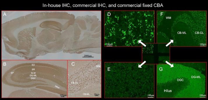
- Nagata N, Kanazawa N, et al. Neuronal surface antigen-specific immunostaining pattern on a rat brain immunohistochemistry in autoimmune encephalitis. Front Immunol. 2023. 13:1066830.