Alzheimer's Disease Study Tools
Alzheimer's disease (AD) is the most common form of dementia and characterized by learning and memory impairment involving extremely complex mechanisms. Creative Bioarray provides a full range of assays which have been extensively practiced in the development of preclinical drugs targeting Alzheimer's disease. Our assays include all kinds of behavioral tests that evaluate changes in general cognitive function of the animals, neurological Imaging that reveals the three-dimensional anatomical structure and function of animal brain, electrophysiology assays that demonstrate neuronal properties (patch-clamp) in brain slices or overall activity (EEG) in animal brain. In addition, we, as always, embrace any newer, updated and advanced techniques developed by the field, and our effort will always be made to incorporate every possible approach to facilitate your research.
 Figure. 1. Neuropathy of dementia
Alzheimer's Disease Study Tools include but not limited to:
- Behavior Tests
- Cognitive Functions Tests
- Sensory Function Tests
- Motor Function Tests
- Social Interaction
- Gait and Balance Changes
- Neurological Imaging
- MRI, PET/CT, and SPECT/CT
- Electrophysiology Assay
- Long-term Potentiation (LTP) Assay
- Patch Clamp
- EEG Monitoring
- Microdialysis
Our extensive experience enables us for any comprehensive tests in Alzheimer's disease research. With professions in customized protocols and assays to precisely meet client’s need, our experts are fully capable and easy to communicate.
Study examples
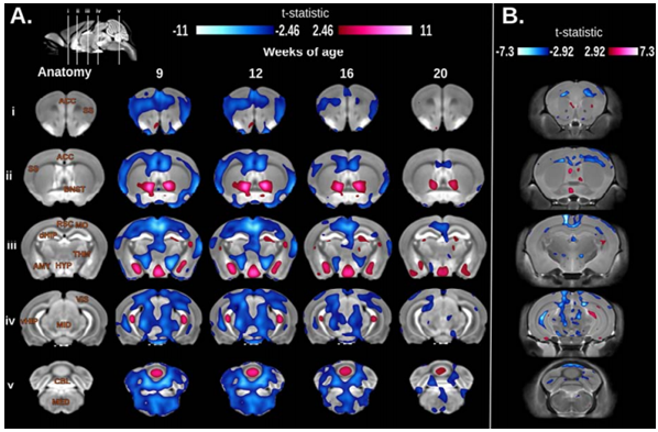 Figure. 2. Longitudinal and cross-sectional voxel-wise differences between TgCRND8 (TG) and WT mice. A. Significant voxel-wise volume differences between TG and WT longitudinally scanned at 9, 12, 16 and 20 weeks of age. Areas of the brain highlighted in blue represent regions where TG mice were significantly smaller than WT at a particular age; red represent regions where TG are significantly larger than WT (t-statistic= ±2.46, 5% FDR). Top left: Sagittal image of the final non-linear average depicting the location of the coronal slice selection through the brain. Rows: Corresponding coronal slices in the rostral to caudal direction (iv). Columns: The first column: Labels of brain regions overlaid on the anatomy (AMY, amygdala; ACC, anterior cingulated cortex; BNST, bed nucleus of the stria terminalis; CBL, cerebellum; dHIP, dorsal hippocampus, vHIP, ventral hippocampus; HYP, hypothalamus; MED, medulla; MID, midbrain; MO, motor cortex; RSC, retrosplenial cortex; SS, somatosensory cortex; THM, thalamus; VIS, visual cortex). Columns 2-5: 9, 12, 16 and 20 weeks of age. B. Results from the cross-sectional volumetric analysis between TG and WT mice at 9, 12, 16, or 20 weeks of age (Blue: TG < WT; Red: TG > WT; t-statistic= ±2.92, 5% FDR)
Quotation and ordering
If you have any special needs or questions regarding our services, please feel free to contact us. We look forward to cooperating with you in the future.
Reference
- Allemang-Grand R et al. Altered brain development in an early-onset murine model of Alzheimer's disease. Neurobiology of Aging. 2015; 36: 638-47.
For research use only. Not for any other purpose.
Disease Models
- Oncology Models
-
Inflammation & Autoimmune Disease Models
- Rheumatoid Arthritis Models
- Glomerulonephritis Models
- Multiple Sclerosis (MS) Models
- Ocular Inflammation Models
- Sjögren's Syndrome Model
- LPS-induced Acute Lung Injury Model
- Peritonitis Models
- Passive Cutaneous Anaphylaxis Model
- Delayed-Type Hypersensitivity (DTH) Models
- Inflammatory Bowel Disease Models
- Systemic Lupus Erythematosus Animal Models
- Oral Mucositis Model
- Asthma Model
- Sepsis Model
- Psoriasis Model
- Atopic Dermatitis (AD) Model
- Scleroderma Model
- Gouty Arthritis Model
- Carrageenan-Induced Air Pouch Synovitis Model
- Carrageenan-Induced Paw Edema Model
- Experimental Autoimmune Myasthenia Gravis (EAMG) Model
- Graft-versus-host Disease (GvHD) Models
-
Cardiovascular Disease Models
- Surgical Models
- Animal Models of Hypertension
- Venous Thrombosis Model
- Atherosclerosis model
- Cardiac Arrhythmia Model
- Hyperlipoidemia Model
- Doxorubicin-induced Heart Failure Model
- Isoproterenol-induced Heart Failure Model
- Arterial Thrombosis Model
- Pulmonary Arterial Hypertension (PAH) Models
- Heart Failure with Preserved Ejection Fraction (HFpEF) Model
- Cardio-Renal-Metabolic (CKM) Syndrome Model
-
Neurological Disease Models
- Alzheimer's Disease Modeling and Assays
- Seizure Models
- Parkinson's Disease Models
- Ischemic Stroke Models
- Acute Spinal Cord Injury (ASCI) Model
- Traumatic Brain Injury (TBI) Model
- Hypoxic-Ischemic Encephalopathy (HIE) Model
- Tourette Syndrome (TS) Model
- Amyotrophic Lateral Sclerosis (ALS) Model
- Huntington's Disease (HD) Model
- Intracerebral hemorrhage (ICH) Models
- Schizophrenia Model
- Depression Models
- Pain Models
-
Metabolic Disease Models
- Type 1 Diabetes Mellitus Model
- Type 2 Diabetes Mellitus Model
- Animal Model of Hyperuricemia
-
Nonalcoholic Fatty Liver Disease Model
- High-Fat Diet-Induced Nonalcoholic Fatty Liver Disease (NAFLD) Model
- Methionine and Choline Deficient (MCD) Diet-Induced Nonalcoholic Fatty Liver Disease (NAFLD) Model
- Gubra-Amylin NASH (GAN) Diet-Induced Nonalcoholic Fatty Liver Disease (NAFLD) Model
- Streptozotocin (STZ) Induced Nonalcoholic Fatty Liver Disease (NAFLD) Model
- High Fat Diet-Induced Obesity Model
- Diabetic Foot Ulcer (DFU) Model
- Cardio-Renal-Metabolic (CKM) Syndrome Model
- Liver Disease Models
- Rare Disease Models
- Respiratory Disease Models
- Digestive Disease Models
-
Urology Disease Models
- Cisplatin-induced Nephrotoxicity Model
- Unilateral Ureteral Obstruction Model
- 5/6 Nephrectomy Model
- Renal Ischemia-Reperfusion Injury (RIRI) Model
- Diabetic Nephropathy (DN) Models
- Passive Heymann Nephritis (PHN) Model
- Adenine-Induced Chronic Kidney Disease (CKD) Model
- Kidney Stone Model
- Doxorubicin-Induced Nephropathy Model
- Orthotopic Kidney Transplantation Model
- Benign Prostatic Hyperplasia (BPH) Model
- Peritoneal Fibrosis Model
- Cardio-Renal-Metabolic (CKM) Syndrome Model
- Orthopedic Disease Models
- Ocular Disease Models
- Infectious Disease Models
- Skin Disease Models
- Otology Disease Models