Anti-Thy1-induced Nephritis Model
Mesangial proliferative glomerulonephritis (MsPGN) is characterized by proliferation of mesangial cells and abnormal extracellular matrix (ECM) accumulation in mesangium. Activated proliferating mesangial cells usually secret large amount of proinflammatory and profibrotic cytokines, including TGF-β, TNF, interleukin-6 (IL-6), and angiotensin II, which are often involved in the pathogenesis of MsPGN.
Anti-Thy 1 nephritis is an experimental model that resemble human mesangial proliferative glomerulonephritis and it is induced by applying antibodies directly against the Thy 1 antigen (CD90) in mesangial cells.
Creative Bioarray, with a prestigious team in the industry, focuses on providing customized experiment services to help customers assess the efficacy of drug candidates and study the associated pathological mechanisms of Type 1 diabetes through this model.
Our capabilities
- We utilize inflammatory bowel disease models to test the efficacy of drug candidates targeting IBD.
- We assess the severity of GN through glomerular damage score system.
- We evaluated the deposition of lg and C3 complement via IHC.
- We evaluate various cytokines through IHC, ELISA, etc.
Assays available
- PK/PD blood collections
- Detection of blood creatinine
- BUN
- Proteinuria
- Histopathological evaluation
- Cell proliferation ability assay
- Immunohistochemistry / Immunofluorescence
With extensive experience in the field of GN, we are confident to help you to overcome any upcoming challenges. Our experts are fully capable of customizing our protocols and assays to meet your specific needs. With our help, we wish to facilitate your research with high efficiency.
Study examples
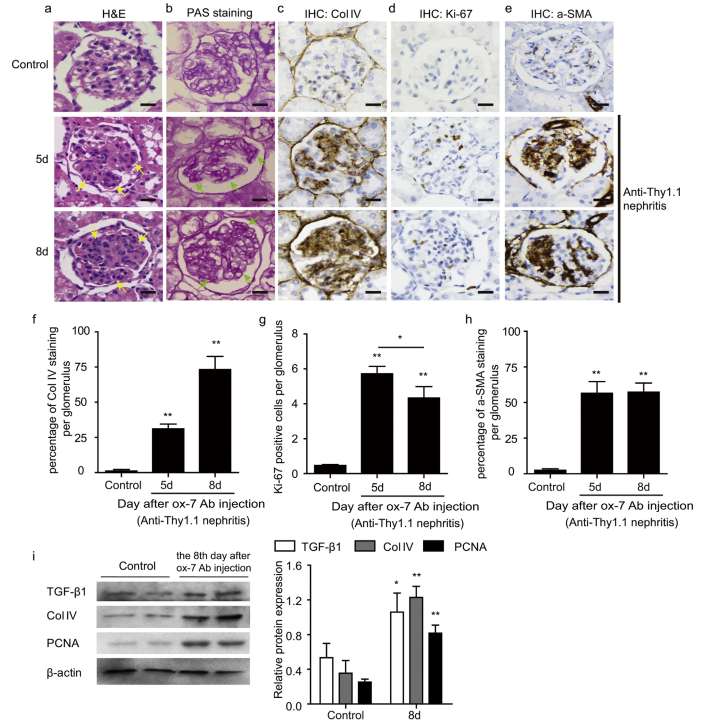 Figure. 1. Rat anti-Thy1.1 nephritis model was induced by ox-7 antibody. (a,b) Representative glomeruli with H&E (a) and PAS staining (b) from Control rats and rats sacrificed on the 5th or 8th day since the injection of ox-7 antibody, showing a progressive cell proliferation and ECM deposit. (c–e) Immunohistochemical staining with Col IV (c), Ki-67 (d) and α-SMA (e) revealed markedly increased collagen accumulation in mesangium and cell proliferation. (f–h) The percentage of Col IV stained area (f), number of Ki-67 positive cells (g) and the percentage of α-SMA stained area (h) per glomerulus were calculated from 25 randomly captured images for each rat (five rats per group) at the original magnification of 400×, showing that they all increased after the injection of ox-7 antibody. As for Ki-67, it peaked at the 5th day in anti-Thy1.1 nephritis and mildly decreased but still elevated at the 8th day, compared to the normal group. (i) Immunoblotting of kidney cortex homogenates indicated up-regulated protein expression of Col IV, TGF-β1 and PCNA in anti-Thy1.1 nephritis rats (day 8 since the injection of ox-7 antibody). Data are expressed as means ± SD, *P < 0.05 for anti-Thy1.1 nephritis rats sacrificed on 5th day vs. 8th day; *P < 0.05 **P < 0.01 for antiThy1.1 nephritis rats vs. control rats. (Scale bar = 100 μm.)
Quotation and ordering
If you have any special needs or questions regarding our services, please feel free to contact us. We look forward to cooperating with you in the future.
Reference
Mao X, Luo W, Sun J, et al. Usp2-69 overexpression slows down the progression of rat anti-Thy1.1 nephritis[J]. Experimental and Molecular Pathology, 2016, 101(2):249-258.