How to Isolate PBMCs from Whole Blood?
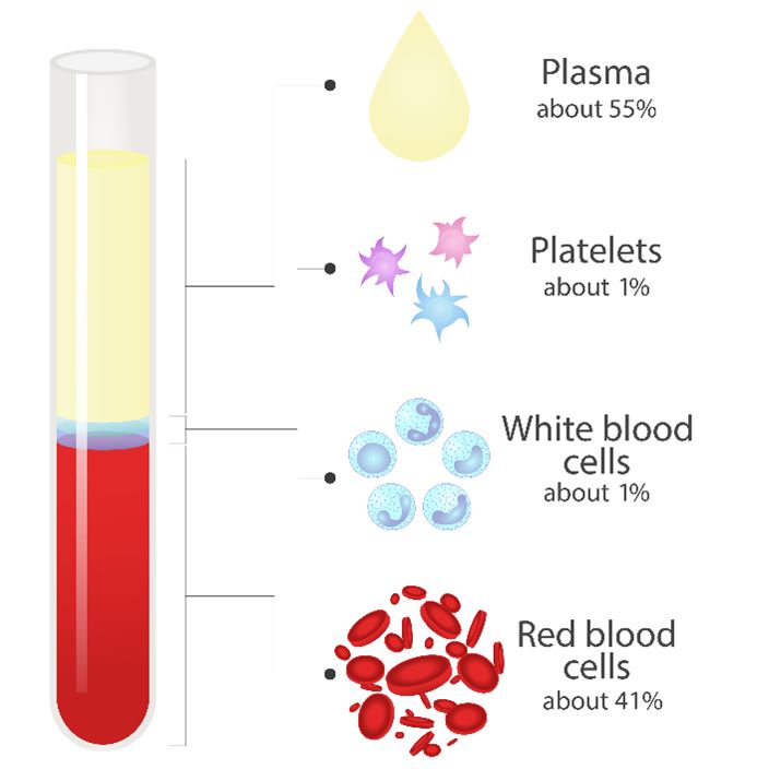
PBMC isolation protocols involve separating lymphocytes directly from whole blood. Whole blood is the blood that flows through the human body — the raw fluid without any components removed or separated. It is mainly composed of plasma, platelets, red blood cells, and PBMCs. PBMCs make up only a small portion (about 1%) of a whole blood sample, and are difficult to study when crowded by other substances. PBMC isolation from whole blood must be performed before researchers can conduct their experiments.
There are six primary methods for isolating PBMCs from whole blood: density-gradient centrifugation, fluorescence activated cell sorting (FACS), magnetic-activated cell sorting (MACS), buoyancy-activated cell sorting (BACS), leukapheresis, and cell preparation tubes (CPT). Each of these methods has its own advantages and disadvantages.
Density-Gradient Centrifugation
Density-gradient centrifugation relies on physical properties such as size and density to sort cell populations. By placing the sample into a centrifuge that rotate at high speed, different types of cells will be sorted out by grouping with particles of a similar density. More dense particles will fall to the bottom or the outside, while less dense particles will stay toward the center or rise to the top. After centrifugation of whole blood, the PBMC layer will appear as a thin white layer underneath the plasma.
Although the process is relatively cheap and simple, density-gradient centrifugation has limited cell specificity, low purity, and low throughput.
Fluorescence-Activated Cell Sorting (FACS)
One technique for immune cell isolation involves the use of a flow cytometer, a machine that uses fluorescent light to label different particles based on characteristics such as size, shape, and radiance. This method, called fluorescence activated cell sorting (FACS), allows samples to flow through a tube and then sorts the components into distinct groups.
FACS requires expensive equipment and properly trained personnel. While this technique is useful when sorting diverse cell samples that need many different populations, it is incredibly time-consuming to use for PBMCs isolation.
Magnetic-Activated Cell Sorting (MACS)
Another method binds magnetic beads to target cells and flows the sample through a magnetic field, suspending the cells with magnets attached. MACS is faster and cheaper than the two previously mentioned methods, but it has the highest cell loss of the three. The harsh magnetic fields can rupture cells, and cause fractured organelles to clutter the sample. This extracellular debris can cause clumping and blockage which results in a lower throughput.
Buoyancy-Activated Cell Sorting (BACS)
BACS leverages the power of gravity to sort cells. This method uses microbubbles that are functionalized to capture analytes of interest and float them to the top of the sample container. The microbubbles are simply mixed into the sample, where they bind to the contaminating cells and float them to the top for removal, leaving the cells of interest at the bottom. The microbubbles carry out their function without any blunt physical forces or harsh magnetic fields, fully maintaining cell health and physiology for downstream applications.
Leukapheresis
Leukapheresis is a procedure that separates blood components to collect specific cells and return the unneeded components back to the circulation. Typically, healthcare professionals use continuous flow centrifugation (CFC) to collect, spin, and return blood continuously while the donor is connected to an instrument called an apheresis circuit. CFC enriches and collects PBMCs by separating whole blood into fractions based on density. After separation, the fractions that do not contain PBMCs are returned to the patient by the apheresis circuit. The main advantage of this system is that only a small volume of blood outside of the donor's circulation during the procedure. Because of this, patients do not typically need fluid replacement during leukapheresis.
Cell Preparation Tubes (CPT)
Sometimes clinical trial protocol specifications are time-critical, and it is more convenient to perform partial on-site processing of PBMCs. To provide faster and easier PBMC isolation, CPT containing anticoagulant plus a gel product will aid in blood layer separation. CPT tubes can be centrifuged on site and then shipped to the laboratory for the final isolation steps. However, if blood quality is poor (e.g., if there is hemolysis or coagulation present) effective PBMC isolation cannot be achieved.
Creative Bioarray Relevant Recommendations
Accelerate your programs with Creative Bioarray's extensive inventory and prospective network of PBMCs.
| Cat. No. | Product Name |
| CSC-7652W | Human Normal Peripheral Blood Mononuclear Cells (PBMCs) |
| CSC-C4085X | Human Peripheral Blood Mononuclear Cells (HPBMC) |
| CSC-00853L | Human iPSC Line from PBMCs |
| CSC-7654W | Human MULTIPLE MYELOMA, Peripheral Blood Mononuclear Cells (PBMCs) |