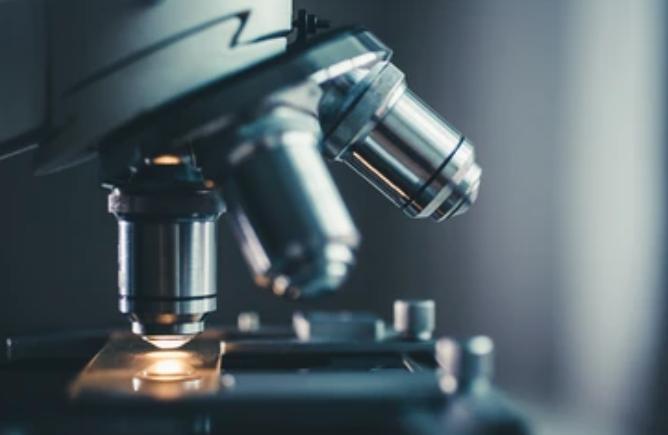
Microscope Platforms

The field of biological imaging has witnessed significant advancements in recent years, thanks to the development of various microscope platforms. These platforms enable scientists and researchers to capture detailed images of biological specimens, uncovering intricate details that were previously inaccessible.
Optical Microscope Platform
The optical microscope platform has long been a staple in biological research due to its versatility and ease of use. Within this platform, several techniques have emerged, revolutionizing the way we observe and understand biological structures.
- Structural illumination microscope
Structural illumination microscopy, also known as SIM, is another technique that has significantly improved optical imaging. By employing patterned illumination and subsequent computational analysis, SIM offers resolutions beyond the diffraction limit. This platform has become invaluable in visualizing cellular structures that demand high-resolution imaging. - Fluorescence lifetime imaging microscope
FILM obtains images based on the differences in the excited state decay rate from a fluorescent sample. As the fluorescence lifetime is not affected by sample concentration, absorption, thickness, photo-bleaching, or excitation intensity, FILM is more robust than intensity-based methods. Besides, as the fluorescence lifetime is dependent on a variety of environmental parameters, FILM can be applied for functional imaging purposes. - Super-resolution optical microscope
Super-resolution optical microscopy has overcome the diffraction limit, allowing researchers to achieve resolutions beyond what was previously possible with traditional light microscopy. By employing techniques such as stimulated emission depletion (STED) microscopy and single-molecule localization microscopy (SMLM), these microscopes can capture images with nanoscale precision.
Electron Microscope Platform
The electron microscope platform offers unparalleled magnification and resolution, making it an indispensable tool for studying the ultrastructure of biological specimens.
- SEM (scanning electron microscope)
Scanning electron microscopy (SEM) allows researchers to capture detailed three-dimensional images of complex biological structures at high magnification. By scanning a focused beam of electrons across the specimen's surface, SEM produces images that showcase intricate details and surface topography. - TEM (transmission electron microscope)
Transmission electron microscopy (TEM) is widely regarded as the gold standard for resolving the finest details in biological specimens. By transmitting electrons through ultrathin sections of samples, TEM creates high-resolution images with extraordinary magnification. Researchers can visualize subcellular structures, such as organelles and macromolecules, and gain valuable insights into their composition and arrangement.
Confocal Microscope Platform
Confocal microscopy has revolutionized biological imaging by eliminating out-of-focus light and enabling researchers to focus on specific cellular structures.
- CLSM (confocal laser scanning microscopy)
Confocal laser scanning microscopy (CLSM) utilizes point illumination and a pinhole aperture, allowing researchers to capture images from a single plane of focus. By rejecting light emitted outside this plane, CLSM produces sharp and highly resolved images of biological samples. - SDCM (spinning disk confocal microscopy)
Spinning disk confocal microscopy (SDCM) is a technique that combines the benefits of confocal imaging with enhanced imaging speed. By utilizing a rotating disk with multiple pinholes, it captures multiple images simultaneously, reducing acquisition times while maintaining superior optical sectioning. SDCM enables researchers to visualize dynamic cellular events, such as live-cell imaging and rapid 3D imaging.
Creative Bioarray Relevant Recommendations
Creative Bioarray has scientists and imaging laboratories to perform the full comprehensive Transmission Electron Microscopy (TEM) and Scanning Transmission Electron Microscopy (STEM) Services for the biological sciences and clinical research including plant samples, animal samples, bacteria, and pathology specimens.