
Renal Proximal Tubule Cells, Secondary, 500,000 cells per vial
Cat.No.: CSC-C4067X
Species: Human
Source: Kidney
Cell Type: Epithelial Cell
- Specification
- Background
- Scientific Data
- Q & A
- Customer Review
Cell Features:
Mixed Renal Epithelial, Renal Cortical Epithelial, and Renal Medullary Epithelial are cryopreserved as primary cells isolated from human kidney tissue and expanded in culture vessels once before cryopreservation.
Renal Proximal Tubule Epithelial are cryopreserved as secondary cells isolated from human kidney tissue and expanded in culture vessels twice before cryopreservation.
All Renal Epithelial Cell types can be grown in a 0.5% serum medium without phenol red or antimicrobials when cultured in LIRen Medium.
All Renal Epithelial Cell types are extensively tested for quality and optimal performance.
Creative Bioarray guarantees performance and quality.
The proximal tubule is composed of Renal Proximal Tubule Cells (RPTCs), the most metabolically active cells in the kidney. They carry out the reabsorption of approximately 65% to 70% of filtered water, electrolytes, and nutrients back from the glomerular filtrate. Given their high metabolic demand and expression of numerous transporters, proximal tubules are the site of greatest vulnerability to nephrotoxic drugs and subsequent acute kidney injury (AKI).
The two main cell lines used in biomedical research today are Human Primary Renal Proximal Tubule Epithelial Cells (hRPTECs) or the immortalized HK-2 cell line. Although HK-2 cells are favored for high throughput studies due to their scalability and reproducibility, hRPTECs are commonly utilized for high-fidelity models because they retain physiological polarization, cilia, and expression of important proximal tubule transporters (OAT1/3, OCT2) better than HK-2 cells.
These cells have been cultured on many cutting-edge in vitro platforms, including Organ-on-a-Chip (OoC) and Microfluidic Biomimetic Chips. Fluid shear stress serves as a stimulus for RPTCs to extend mature microvilli and tightly attach to the basement membrane (i.e. through Na/K-ATPase expression). As a result, RPTECs are being used to screen drugs, study metabolism, and model kidney diseases like diabetic nephropathy and fibrosis.
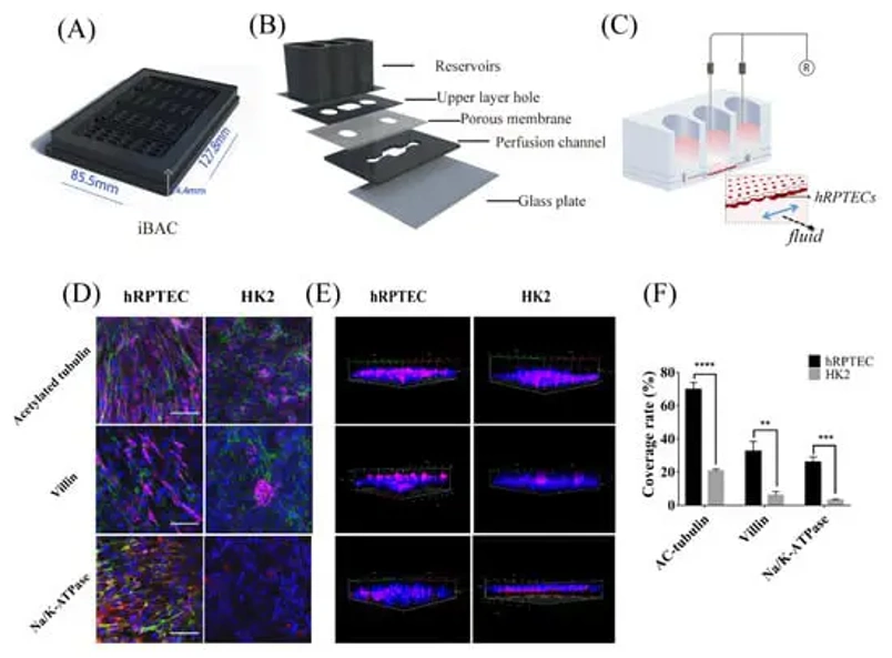
Functional Evaluation and Nephrotoxicity Assessment of Human Renal Proximal Tubule Cells on a Chip
For clinically relevant prediction of nephrotoxicity it is critical to model native transporter expression and epithelial polarization in vitro. Here, Jing's team sought to establish and characterize a high-throughput model of the renal proximal tubule using an integrated biomimetic array chip (iBAC).
Resembling the footprint of a conventional 96-well plate (Fig. 1A), the iBAC consists of 24 individual units arrayed together. Within each unit there are five layers comprising a 0.4 µm PET porous membrane for apico-basal permeability and perfusion channel that enables application of physiologically relevant biomimetic fluid shear stress (Fig. 1B). Primary human renal proximal tubule epithelial cells (hRPTECs) or immortalized HK2 cells were seeded on the basal side of the membrane and allowed to adhere (Fig. 1C). Shear stress magnitude and enhancement of nutrient supply were controlled by placing the iBAC on an angular shaker with user-defined angle and frequency settings.
Cells remained adherent on the device for greater than one week. Polarization of hRPTECs or HK2 cells was compared after dynamic culture for seven days. Immunofluorescence staining of acetyl-α-tubulin (cilia) and villin (microvilli) showed significantly greater apical protein expression on hRPTECs relative to HK2 cells (Fig. 1D, E). Furthermore, hRPTECs exhibited significantly greater basal expression of sodium reabsorption enzyme Na/K-ATPase (Fig. 1D, E). Quantification of percent protein coverage showed that hRPTECs undergo a more mature/polarized phenotype relative to HK2 cells after seven days in culture on the iBAC (Fig. 1F).
Ask a Question
Write your own review
Description: Renal glomerular endothelial cells (GEC) are a specialized microvascular cell type involved in the regulation of glomerular ultrafiltration. They form the inner part of the filtration barrier and are ...
Description: Human Kidney Endothelial Cells from Creative Bioarray are isolated from human kidney tissue. Human Kidney Endothelial Cells are grown in T25 tissue culture flasks pre-coated with gelatin-based ...
Description: Human Proximal Tubular Epithelial Cells from Creative Bioarray are isolated from normal human proximal tubular tissue. Human Proximal Tubular Epithelial Cells are grown in T25 tissue culture flasks ...
Description: Primary Human Glomerular Microvascular Endothelial Cells were initiated by decapsulated glomeruli isolated from normal human kidney cortical tissue.These cells were originated using Complete ...
Description: Human Kidney Carcinoma Epithelial Cells from Creative Bioarray are isolated from human kidney tumor tissue. Human Kidney Carcinoma Epithelial Cells are grown in T25 tissue culture flasks pre-coated ...
Description: VE-Cad-GFP Expressing Human Glomerular Microvascular Endothelial Cells (VE-Cad-GFP HGMvECs) provided by Creative Bioarray are puromycin-selected from Human Glomerular Microvascular Endothelial Cells ...