
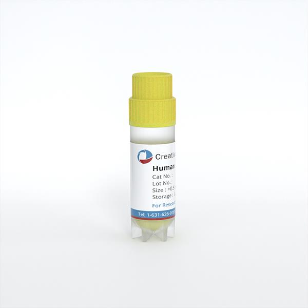
Human Glomerular Microvascular Endothelial Cells
Cat.No.: CSC-C4379X
Species: Human
Source: Kidney
Cell Type: Endothelial Cell; Microvascular Cell
- Specification
- Background
- Scientific Data
- Q & A
- Customer Review
These cells were originated using Complete Serum-Free Medium Kit With SuperFuel™, are available at <12 Cumulative Population Doublings (CPD) in vitro [Passage 3] and were cryopreserved in aliquots of ~1.5 X 10^6 cells. This vial will initiate a Passage 4 cell culture in a 75cm2 flask.
Human Glomerular Microvascular Endothelial Cells are specialized endothelial cells lining the capillaries of renal glomeruli. Together with the glomerular basement membrane and podocytes, they form the glomerular filtration barrier. In vivo, their fenestrated structure supports high water and small-solute permeability, while the endothelial glycocalyx contributes to restricting plasma protein passage. This combination makes them particularly relevant to studies of renal filtration and glomerular vascular integrity.
Their principal advantage is the combination of human origin and glomerular tissue specificity. They enable direct investigation of the endothelial contribution to kidney function in a human cellular context. Appropriately characterized cultures support measurements of albumin permeability, junctional organization, glycocalyx integrity, and responses to vascular regulatory signals.
These cells also provide a valuable platform for investigating glomerular injury. Human glomerular endothelial studies have shown that high glucose can impair glycocalyx-dependent barrier function, while inflammatory stimulation can alter permeability and cellular morphology. Such responses support mechanistic research into diabetic kidney disease, inflammatory endothelial injury, and pathways contributing to proteinuria, as well as evaluation of candidate endothelial protective interventions.
An additional strength is their integration into podocyte co-cultures and glomerulus-on-a-chip systems. Incorporating mesangial cells, extracellular matrix, and controlled flow enables investigation of multicellular interactions and improves the physiological relevance of filtration models.
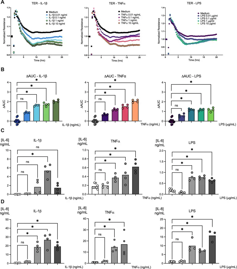
Microbial and Host Inflammatory Factors Induce Permeability and Activate Cytokine Production by Human Glomerular Microvascular Endothelial Cells
The broad impacts of microbial and endogenous inflammatory agonists on the promotion of systemic inflammation and on some vascular endothelial cells have been extensively characterized. Furthermore, endothelial dysfunction is a key feature of sepsis and may contribute to acute kidney injury (AKI). However, the role of glomerular endothelial cells in the pathophysiology of sepsis-associated AKI is not well characterized. To address this gap, we tested the effects of bacterial LPS, and the cytokines, IL-1β and TNF-α, on the barrier function and IL-6/IL-8 production of human glomerular microvascular endothelial cells (HGMVEC).
The effects of IL-1β, TNF-α, and LPS on the barrier function of HGMVEC were assessed using TER as a surrogate for permeability. Compared to treatment with cell culture medium, treatment with each inflammatory agonist induced a concentration-dependent reduction in TER across all tested concentrations over 24 h (Fig. 1A, 1B). These results indicated that all three inflammatory agonists induced HGMVEC permeability.
We quantified IL-6 and IL-8 levels in HGMVEC culture supernatants after 24 h. Compared with the medium, treatment with IL-1β, TNF-α, and LPS significantly increased the production of IL-6 (Fig. 1C) and IL-8 (Fig. 1D) in a concentration-dependent manner. Of the three inflammatory agonists, IL-1β exhibited the highest potency in inducing IL-6 and IL-8 production (EC50 0.0134 ng/mL) by HGMVEC, followed by LPS (EC50 0.179 ng/mL) and TNF-α (EC50 1.31 ng/mL). In conclusion, this study demonstrates that cytokines and microbial LPS, each robustly induce IL-6 and IL-8 and hyperpermeability in HGMVEC.
Ask a Question
Write your own review
Description: Renal glomerular endothelial cells (GEC) are a specialized microvascular cell type involved in the regulation of glomerular ultrafiltration. They form the inner part of the filtration barrier and are ...
Description: Human Kidney Endothelial Cells from Creative Bioarray are isolated from human kidney tissue. Human Kidney Endothelial Cells are grown in T25 tissue culture flasks pre-coated with gelatin-based ...
Description: Human Proximal Tubular Epithelial Cells from Creative Bioarray are isolated from normal human proximal tubular tissue. Human Proximal Tubular Epithelial Cells are grown in T25 tissue culture flasks ...
Description: The adrenal cortex makes up the perimeter of the adrenal gland and plays an essential role in regulating homeostasis in the body through the secretion of corticosteroid and androgen hormones. The ...
Description: Human Kidney Glomerular Endothelial Cells from Creative Bioarray are isolated from human kidney tissue. Human Kidney Glomerular Endothelial Cells are grown in T25 tissue culture flasks pre-coated ...