
Human Keratocytes (HK)
Cat.No.: CSC-7765W
Species: Human
Source: Cornea; Eye
Cell Type: Keratocyte
- Specification
- Background
- Scientific Data
- Q & A
- Customer Review
Human Keratocytes (HK) are a primary human cell culture that is used as model cells to study the cornea in vitro. HK are neural crest derived mesenchymal cells (commonly referred to as corneal fibroblasts) located in the stroma between lamellae of collagen arranged perpendicularly. They synthesize collagen and proteoglycans of the corneal stroma.
HK cells maintain a typical fibroblast spindle shape in culture and stain positive for fibronectin. They are generally frozen as primary culture or passage one. Cell banks can assure that these cells will grow 12-15 population doublings in fibroblast growth medium supplemented with low serum and growth factors. HK can phenotypically change from their resting state to an active repair state upon stimulation with cytokines. HK express the interleukin receptor proteins IL-4R and IL-17R. They are used to study wound healing in the cornea, development of fibrosis, drug permeability, and regenerative medicine to treat corneal disorders of the stroma.
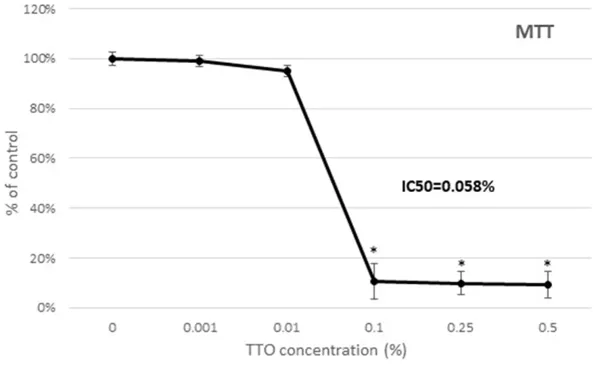
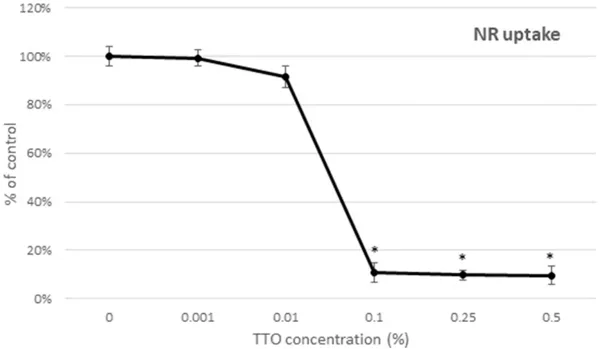
Tea Tree Oil Influence on Human Keratocytes Growth and Viability
Tea tree oil (TTO) is used in ophthalmology for eyelid health and treating parasitic infections, but excessive exposure may harm corneal keratocytes-cells essential for maintaining corneal homeostasis. Paduch et al. aimed to determine safe TTO concentrations for these cells using a normal human keratocyte (HK) cell line.
Cell viability analysis was performed using the MTT method (Fig. 1) and NR uptake assay (Fig. 2). Data obtained using both methods indicated a similar profile of cytotoxic activity of tea tree oil (TTO) on keratocytes. TTO concentrations up to 0.01% had no toxic activity on HK cells. Cell metabolic activity and thus indirectly viability was 94.9% (Fig. 1) and viability 91.6% (Fig. 2) at TTO concentration 0.01% as compared to control. The use of concentrations exceeding 0.01% significantly reduced cell viability. At the concentrations 0.1%; 0.25% and 0.5% of TTO the viability of HK cells were significantly reduced to about 10% as compared to control (Fig. 1, Fig. 2). The IC50 values were 0.058% obtained by the MTT method and 0.056% calculated by the NR uptake assay.
Ask a Question
Write your own review

Description: Human choroidal endothelial cells are isolated from normal human choroidal tissue. The cells are provided at passage 1 and shipped in frozen vials. These cells have a minimum average population ...
Description: Human Non-Pigment Ciliary Epithelial Cells (HNPCEpiC) from Creative Bioarray are isolated from human ciliary bodies. HNPCEC are cryopreserved at primary culture and delivered frozen. Each vial ...
Description: RFP Expressing Human Retinal Microvascular Pericytes (RFP-HRMPs) provided by Creative Bioarray are zeocin-selected from Human Retinal Microvascular Pericytes infected with lentivirus expressing RFP. ...

Description: Primary Human Retinal Pericyte Cells (HRPEpiC) are isolated from human retina. HRPEpiC are cryopreserved at passage one and delivered frozen. Each vial contains >5 x 10^5 cells in 1 ml volume. ...
Description: Human Retinal Pigment Epithelial Cells (HRPEpiC) are isolated from human retina. HRPEpiC are cryopreserved at passage one and delivered frozen. Each vial contains >5 x 10^5 cells in 1 ml volume. ...
Description: Human Conjunctival Fibroblasts (HConF) from Creative Bioarray are isolated from human conjunctiva. HConF are cryopreserved at primary culture and delivered frozen. Each vial contains >5 x 10^5 cells ...