
Human Non-Pigment Ciliary Epithelial Cells (HNPCEpiC)
Cat.No.: CSC-7750W
Species: Human
Source: Ciliary Body; Eye
Cell Type: Epithelial Cell
- Specification
- Background
- Scientific Data
- Q & A
- Customer Review
Human Non-Pigmented Ciliary Epithelial Cells (HNPCEpiC) are primary epithelial cells derived from the non-pigmented layer of the human ciliary body epithelium. The ciliary epithelium is composed of pigmented and nonpigmented epithelial layers that cooperate to control the generation and content of aqueous humor. Among these, non-pigmented ciliary epithelial cells are the key players in aqueous humor secretion with active ion transportation and osmotic regulation, therefore contributing to the maintenance of intraocular pressure (IOP) and ocular homeostasis.
HNPCEpiC displayed classic epithelial shape and expressed transporters, ion channels, and tight junction proteins involved in fluid transport and barrier function. These cells offer a physiologically relevant in vitro model for studying aqueous humor dynamics, ciliary body physiology and processes regulating intraocular pressure. In addition, recent studies have revealed shown the existence of primary cilia and mechanosensitive signaling pathways in HNPCE cells, emphasizing their importance in pressure sensing and glaucoma research.
Human non-pigmented ciliary epithelial cells are commonly utilized in studies of glaucoma, ocular hypertension, aqueous humor secretion, ion transport, mechano-transduction and blood-aqueous barrier function. They are also useful in the evaluation of ophthalmic medication candidates, which are intended to modulate the production of aqueous humor and the regulation of intraocular pressure. HNPCEpiC as a biologically realistic ocular cell model provide a significant platform for the study of eye physiology, disease processes and development of innovative therapeutic strategies for glaucoma and other anterior segment problems.
TRPP2 Expression and Serum Starvation–Induced Primary Cilia Formation in Human Non-Pigmented Ciliary Epithelial Cells
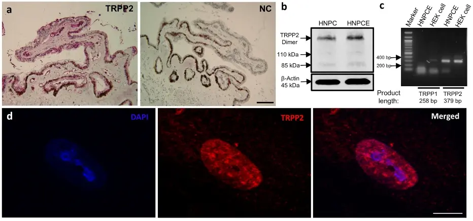
Mechanosensitive channels and primary cilia may sense intraocular pressure (IOP), but their expression in the ciliary body epithelium (CBE) is poorly characterized. Zheng’s team examined TRPP2 and primary cilia in a human non‑pigmented ciliary epithelial (HNPCE) cell line.
Immunohistochemistry confirmed TRPP2 expression in both non‑pigmented and pigmented CBE cells in human ocular tissue. HNPCE cultures likewise expressed TRPP2, verified by Western blot, PCR, and immunofluorescence (Fig. 1b–d). TRPP2 localized predominantly to the nucleus and showed a punctate cytoplasmic distribution.
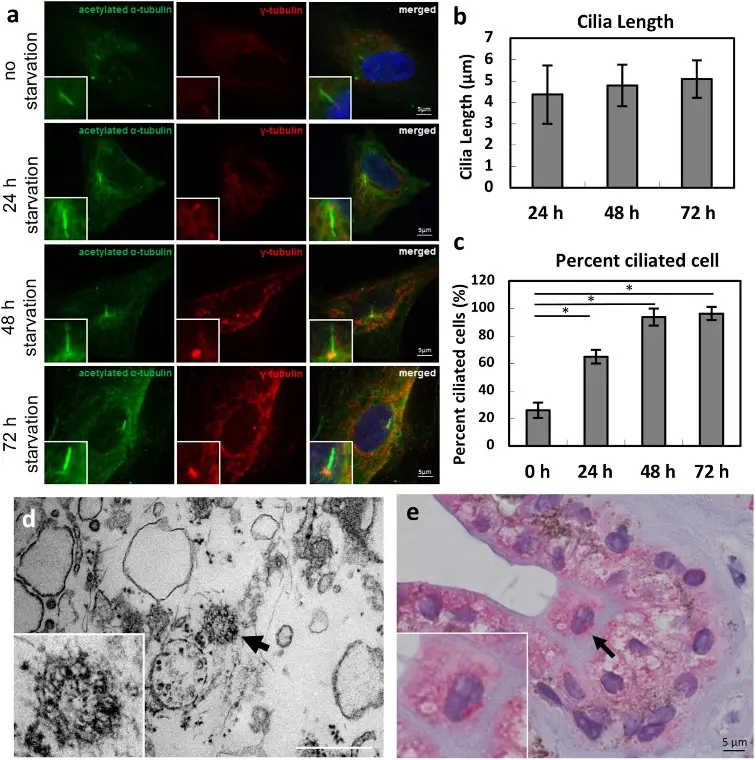
Primary cilia were detected using acetylated α‑tubulin (axoneme) and γ‑tubulin (basal body) markers (Fig. 2a). Cilia length remained stable over serum starvation (24 h: 4.4 ± 1.4 µm; 48 h: 4.8 ± 1.0 µm; 72 h: 5.1 ± 0.9 µm; p= ns) (Fig. 2b). In contrast, the percentage of ciliated cells increased markedly with starvation: 26.0 ± 5.6% (0 h), 65.0 ± 4.9% (24 h), 93.9 ± 6.1% (48 h), and 96.4 ± 4.7% (72 h) (Fig. 2c).
These results demonstrate TRPP2 expression in HNPCE cells and show that serum starvation robustly induces primary cilia formation without affecting cilia length—providing a foundation to study mechano-sensation in the ciliary body.
Human Non-Pigment Ciliary Epithelial Cells (HNPCEpiC) from Creative Bioarray are guaranteed to further expand for 10 population doublings.
Ask a Question
Write your own review
Description: Human choroidal endothelial cells are isolated from normal human choroidal tissue. The cells are provided at passage 1 and shipped in frozen vials. These cells have a minimum average population ...
Description: Human Keratocytes (HK) from Creative Bioarray are isolated from human cornea. HK are cryopreserved at primary culture. Each vial contains >5 x 10^5 cells in 1 ml volume. HK are characterized by their ...
Description: RFP Expressing Human Retinal Microvascular Pericytes (RFP-HRMPs) provided by Creative Bioarray are zeocin-selected from Human Retinal Microvascular Pericytes infected with lentivirus expressing RFP. ...
Description: Primary Human Retinal Pericyte Cells (HRPEpiC) are isolated from human retina. HRPEpiC are cryopreserved at passage one and delivered frozen. Each vial contains >5 x 10^5 cells in 1 ml volume. ...
Description: Human Retinal Pigment Epithelial Cells (HRPEpiC) are isolated from human retina. HRPEpiC are cryopreserved at passage one and delivered frozen. Each vial contains >5 x 10^5 cells in 1 ml volume. ...
Description: Human Conjunctival Fibroblasts (HConF) from Creative Bioarray are isolated from human conjunctiva. HConF are cryopreserved at primary culture and delivered frozen. Each vial contains >5 x 10^5 cells ...