
Porcine Preadipocytes
Cat.No.: CSC-C10057
Species: Pig
Source: Adipose
Cell Type: Preadipocyte
- Specification
- Background
- Scientific Data
- Q & A
- Customer Review
Porcine preadipocytes are mesenchymal progenitor cells derived from the stromal vascular fraction of adipose tissue depots. These cells exhibit a characteristic fibroblast-like, spindle-shaped morphology in monolayer culture, with robust proliferative capacity prior to commitment. Upon adipogenic induction with a cocktail of insulin, dexamethasone, 3-isobutyl-1-methylxanthine (IBMX), and a PPARγ agonist, porcine preadipocytes terminally differentiate into mature adipocytes, as evidenced by prominent lipid droplet accumulation and upregulated expression of canonical markers including PPARγ, C/EBPα, FABP4 (aP2), and perilipin.
A primary advantage of porcine preadipocytes lies in the exceptional translational relevance of the pig model. Pigs share striking similarities with humans in cardiovascular physiology, metabolic pathways, lipoprotein profiles, and adipose tissue distribution, rendering them superior to rodents for studying obesity, insulin resistance, type 2 diabetes, and metabolic syndrome. Porcine preadipocytes retain depot-specific heterogeneity, with intramuscular preadipocytes displaying distinct transcriptomic profiles and adipogenic capacities compared to subcutaneous counterparts-a feature critical for investigating marbling fat deposition, which directly impacts pork quality and consumer preference.
Furthermore, porcine preadipocytes are indispensable for agricultural biotechnology, enabling research into nutritional regulation, feed additives, and genetic improvement of meat quality. They also serve as a robust platform for evaluating pharmaceutical candidates targeting lipid metabolism and inflammation.
Oleic Acid and Transferrin Synergistically Induce Serum-Free Adipogenic Differentiation of Porcine Preadipocytes
Cultured meat represents an emerging frontier in cellular agriculture, garnering increasing interest due to its potential benefits regarding sustainability, animal welfare, and food safety. However, its development is hampered by challenges in flavor modulation and sensory quality, primarily due to the limited biosynthesis of fat-derived flavor compounds. Although adipose tissue engineering has been extensively studied, its industrial-scale production is hampered by serum dependency and low differentiation efficiency. Therefore, the establishment of serum-free, efficient strategies for regulating lipid synthesis is urgently needed.
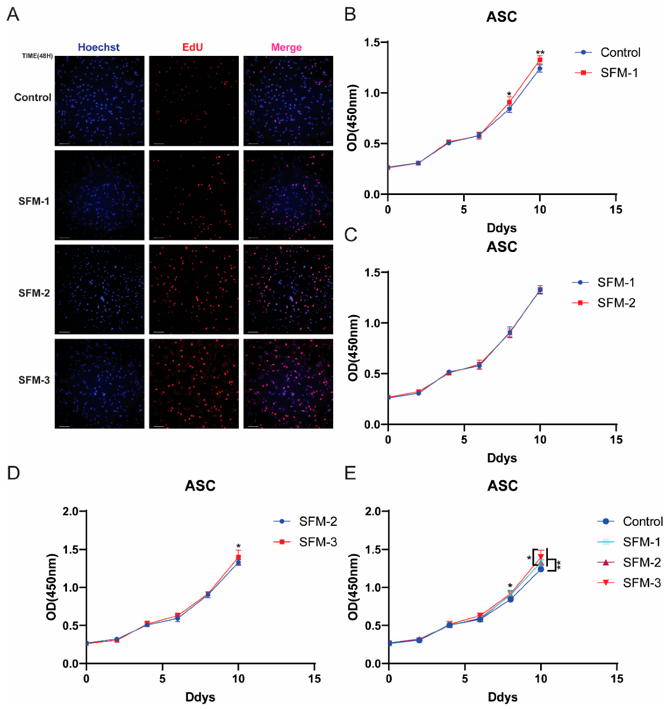
In this study, Fu, Zhou, et al. developed a serum-free adipogenic induction system and investigated its underlying regulatory mechanisms. They demonstrated that Serum-Free Differentiation Medium 1 (SFM-1) initiated the differentiation program and induced intracellular lipid deposition in preadipocytes (~10% by Day 8). Serum-free differentiation medium 2 (SFM-2), which supplied oleic acid (OA) as a lipid substrate and signaling activator, markedly enhanced lipid droplet accumulation and differentiation efficiency. Ultimately, serum-free differentiation medium 3 (SFM-3), leveraging the synergistic action of oleic acid (OA) and transferrin (TRF), successfully activates the expression of SEPTIN4, which in turn regulates a core adipogenic network-including the master transcription factors PPARγ and CEBPα, as well as downstream functional genes. Mechanistically, the OA/TRF combination in SFM-3 upregulates SEPTIN4, unveiling a previously unrecognized regulatory axis that activates the PPARγ signaling pathway, thereby synchronizing the proliferation and differentiation of precursors and guiding them from initiation to functional maturity.
Ask a Question
Write your own review
Description: Porcine Corneal Epithelial Cells from Creative Bioarray are isolated from corneal tissue of porcine. Porcine Corneal Epithelial Cells are grown in a T25 tissue culture flask pre-coated with ...
Description: Porcine Brain Vascular Fibroblasts from Creative Bioarray are isolated from brain tissue of porcine. Porcine Brain Vascular Fibroblasts are grown in T75 tissue culture flasks pre-coated with ...
Description: Porcine Thymus Endothelial Cells from Creative Bioarray are isolated from thymus tissue of porcine. Porcine Thymus Endothelial Cells are grown in T25 tissue culture flasks pre-coated with ...
Description: Porcine Kidney Endothelial Cells from Creative Bioarray are isolated from kidney tissue of porcine. Porcine Kidney Endothelial Cells are grown in T25 tissue culture flasks pre-coated with ...
Description: Pig bone marrow from Creative Bioarray is procured from the fresh femurs. Bone Marrow-CD34+ stem/progenitor cells are positively isolated using a direct immunomagnetic CD34 MicroBead labeling system.
Description: Porcine Primary Thymus Fibroblasts from Creative Bioarray are isolated from thymus tissue of porcine. Porcine Primary Thymus Fibroblasts are grown in T75 tissue culture flasks pre-coated with ...