

Porcine Preadipocyte Cells
Cat.No.: CSC-C0522Z
Species: Pig
Source: Adipose
Morphology: Polygonal
Culture Properties: Adherent
Cell Type: Preadipocyte
- Specification
- Background
- Scientific Data
- Q & A
- Customer Review
Note: Never can cells be kept at -20°C.
Porcine Preadipocyte Cells are primary precursor cells derived from pig adipose tissue, and are capable of adipogenic differentiation under adequate culture conditions. These cells are immature adipocyte precursors and are an early stage of adipose tissue development. Preadipocytes undergo coordinated changes in cell shape, gene expression and fat accumulation during adipogenesis, associated with activation of important regulatory factors such as PPARγ and C/EBP family proteins. They have been utilized as a model to study the molecular pathways that regulate adipocyte differentiation, adipose tissue expansion and metabolic control.
Porcine preadipocyte cells are often utilized in research of obesity, energy metabolism, insulin sensitivity, and adipose derived signaling because of physiological similarities between porcine and human adipose tissue. They may also be used as an experimental model to evaluate the effect of nutrition, hormones, bioactive molecules and genetic variables on the adipogenic processes. Moreover, the cells are used in cattle growth, meat quality improvement and comparative adipose biology researches.
ADAR1 Promotes Proliferation of Porcine Preadipocytes Through Regulation of Cell Cycle
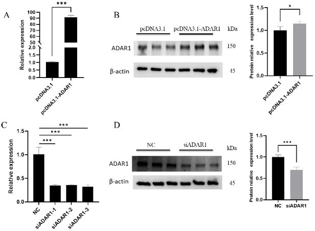
Adenosine deaminase acting on RNA 1 (ADAR1) is an essential regulator of fat metabolism, which affects productivity of cattle and human health. Here, Yang's team examined the function of ADAR1 in porcine preadipocytes using over-expression and knockdown techniques. Transfection with pcDNA3.1-ADAR1 resulted in an increase of ADAR1 mRNA levels by more than 90-fold and a considerable rise in protein expression (Fig. 1A, 1B). In contrast, siADAR1-3 showed the best knockdown efficacy, dropping mRNA by >65% and protein levels (Fig. 1C, 1D).
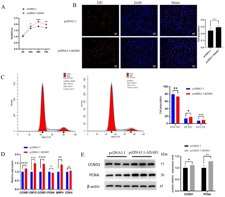
Functional experiments showed that the overexpression of ADAR1 significantly increased the cell survival at 24, 48 and 72 hours post-transfection compared to the empty vector control (Fig. 2A). In line with this, EdU incorporation experiments showed a significant increase in the number of proliferating cells (Fig. 2B). Flow cytometry analysis showed that overexpression of ADAR1 decreased the percentage of cells in G1 phase and increased the percentages of cells in S and G2 phases, which suggests an acceleration of cell cycle progression (Fig. 2C). These conclusions were confirmed by molecular analysis. RT-qPCR indicated a considerable elevation of the major proliferation markers CCNE1, CCND1, CMYC, BMP4 and CDK4 (Fig. 2D). Western blotting also verified the elevated protein levels of CCND1 and proliferative marker PCNA (Fig. 2E). Together, these data suggest that ADAR1 enhances porcine preadipocyte proliferation by promoting cell cycle progression and key genes related to proliferation.
The most common cause is due to denaturation of lipoproteins in the serum, while hemofibrin (one of the proteins that form coagulation) is also present in the serum after it has been thawed and is one of the main causes of precipitates.
Ask a Question
Average Rating: 5.0 | 1 Scientist has reviewed this product
Easy success
Following the cell culture method as described in the instructions, we achieved easy experimental success.
02 Aug 2023
Ease of use
After sales services
Value for money
Write your own review
Description: Porcine Corneal Epithelial Cells from Creative Bioarray are isolated from corneal tissue of porcine. Porcine Corneal Epithelial Cells are grown in a T25 tissue culture flask pre-coated with ...
Description: Porcine Brain Vascular Fibroblasts from Creative Bioarray are isolated from brain tissue of porcine. Porcine Brain Vascular Fibroblasts are grown in T75 tissue culture flasks pre-coated with ...
Description: Porcine Thymus Endothelial Cells from Creative Bioarray are isolated from thymus tissue of porcine. Porcine Thymus Endothelial Cells are grown in T25 tissue culture flasks pre-coated with ...
Description: Porcine Kidney Endothelial Cells from Creative Bioarray are isolated from kidney tissue of porcine. Porcine Kidney Endothelial Cells are grown in T25 tissue culture flasks pre-coated with ...
Description: Pig bone marrow from Creative Bioarray is procured from the fresh femurs. Bone Marrow-CD34+ stem/progenitor cells are positively isolated using a direct immunomagnetic CD34 MicroBead labeling system.
Description: Porcine Primary Thymus Fibroblasts from Creative Bioarray are isolated from thymus tissue of porcine. Porcine Primary Thymus Fibroblasts are grown in T75 tissue culture flasks pre-coated with ...