
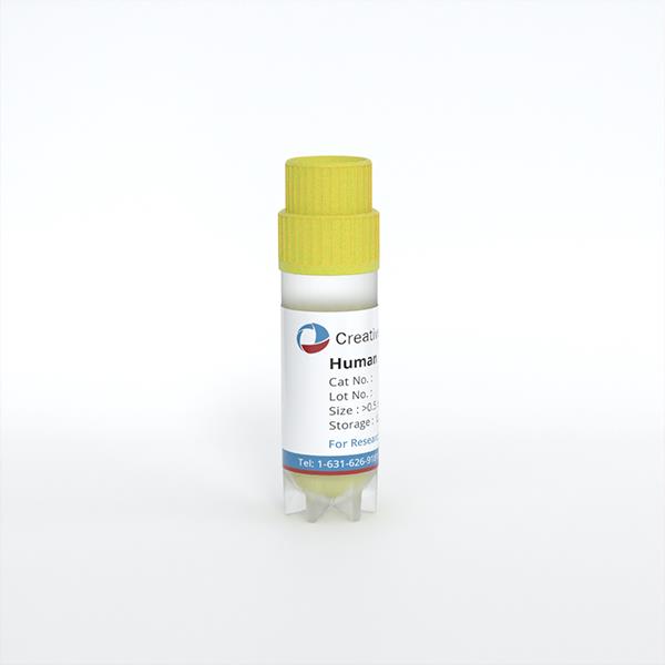
Human Preadipocytes - Visceral (HPAs-v)
Cat.No.: CSC-C4009X
Species: Human
Source: Adipose
Cell Type: Preadipocyte
- Specification
- Background
- Scientific Data
- Q & A
- Customer Review
Human Preadipocytes - Visceral (HPAs-v) from Cretive Bioarray are isolated from human visceral fat tissue. HPA-v are cryopreserved at passage one and delivered frozen. Each vial contains at least 0.5 x 10^6 cells in 1 ml volume. HPAs-v are negative for HIV-1, HBV, HCV, mycoplasma, bacteria, yeast and fungi. HPAs-v are guaranteed for further culture under conditions specified by Creative Bioarray.
Human visceral preadipocytes (HPAs-v) are primary mesenchymal progenitor cells isolated from the stromal vascular fraction of omental adipose tissue. As non-immortalized, diploid cells derived directly from human donors, they retain the complete depot-specific molecular identity of intra-abdominal fat, offering a physiologically authentic system that is unconfounded by the species-dependent metabolic divergences inherent to rodent models.
The defining advantage of HPAs-v is their faithful recapitulation of the pathogenic hallmarks of visceral obesity: they exhibit high basal and catecholamine-stimulated lipolytic rates coupled with profound resistance to the anti-lipolytic action of insulin, thereby replicating the excessive free fatty acid flux that drives ectopic lipid deposition and hepatic insulin resistance. In parallel, they constitutively secrete a pro-inflammatory panel-including elevated IL-6, TNF-α, and MCP-1-while producing disproportionately low adiponectin, thus mirroring the chronic low-grade inflammation that characterizes visceral adipose tissue dysfunction.
HPAs-v display a uniquely heightened sensitivity to glucocorticoids due to abundant expression of the glucocorticoid receptor and the enzyme 11β-hydroxysteroid dehydrogenase type 1, making them an exquisite model for studying cortisol-driven central adipogenesis and its metabolic consequences. They are amenable to lentiviral gene delivery and siRNA-mediated knockdown, facilitating mechanistic gene-function analyses, and can be co-cultured with macrophages to dissect paracrine inflammatory crosstalk.
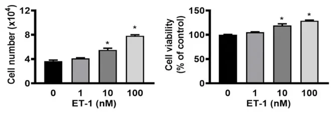
Endothelin-1 Stimulates the Growth of Visceral and Subcutaneous Human Preadipocytes
Endothelin-1 (ET-1) regulates adipogenesis and the endocrine activity of adipocytes in obesity. Human white adipocytes are central to energy storage and endocrine regulation. However, relatively little is known about the involvement of the ET-1 signaling pathway in the growth of human white preadipocytes (HWPs). Therefore, this study investigated the cellular signaling mechanisms in HWPs, focusing on the cellular and functional basis of the actions of ET-1.
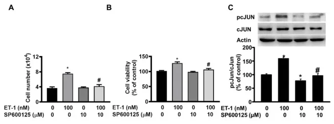
In this study, signaling protein levels, cell proliferation, and the numbers of visceral HWPs were measured by immunoblotting and MTT, and trypan blue exclusion assays. Both ET type A receptor (ETAR) and ET type B receptor (ETBR) antagonists inhibited the ET-1-induced growth of visceral HWPs and the phosphorylation of AMPK, PKC, and STAT3 in these cells. The ETBR antagonist alone blocked the ET-1-induced phosphorylation of ERK and c-JUN. Pretreatment with specific inhibitors of AMPK, ERK, JNK, STAT3, and PKC prevented ET-1-induced cell proliferation and attenuated the phosphorylation of AMPK, ERK, c-JUN, STAT3, and PKC induced by ET-1. These distinct signaling pathways, along with the optimization of pathway-specific inhibitors, have potential implications for the future management of obesity and metabolic disorders.
Ask a Question
Write your own review
Description: 3T3-L1 murine adipocytes have been fundamental in metabolic disease research for over 30 years. Originally derived from Swiss mouse embryo tissue by Dr. Howard Green of Harvard Medical School, the ...
Description: Diseased Preadiopcytes are isolated from diabetic type II donors. Further donor information may be available including BMI, medications taken, and disease duration.Preadipocytes are isolated from ...
Description: Visceral preadipocytes can be cultured as growing precursor cells or differentiated into adipocytes using medium supplemented with adipogenic and lipogenic hormones. This instruction manual describes ...
Description: Preadipocytes are isolated from omental, mesenteric, or perirenal adipose tissue and are tested for their ability to differentiate in culture to mature adipocytes. Preadipocytes are available ...
Description: Diseased Preadiopcytes are isolated from diabetic type I or type II donors. Further donor information may be available including BMI, medications taken, and disease duration.Preadipocytes are ...
Description: Microvascular endothelial cells line blood vessels and contribute to many biological processes such as angiogenesis, coagulation, trafficking of lymphocytes, and the inflammatory response. ...