
Human Preadipocytes, subcutan Cells-Diabetes Type II
Cat.No.: CSC-C8012L
Species: Human
Source: Adipose
Cell Type: Preadipocyte
- Specification
- Background
- Scientific Data
- Q & A
- Customer Review
Preadipocytes are isolated from subcutaneous or visceral fat. Subcutaneous fat is often found attached to the skin in the lower abdomen area. Visceral fat is associated with internal organs such as the bladder and kidney. Relative to subcutaneous fat, visceral fat deposits are mobilized at a higer rate to produce fatty acids and other related cardiovascular disorders. Preadipocytes are precursor cells that develop into adipocytes when fully differentiated. Adipocytes performs essential functions of energy metabolism and are characterized by the accumulation of intracellular triglycerides.
Human Preadipocytes, Subcutaneous Cells – Diabetes Type II are primary adipocyte precursor cells derived from the subcutaneous adipose tissue of donors with Diabetes Mellitus, Type 2 (T2DM). These cells offer an important in vitro model for the study of the cellular and molecular pathways involved in obesity-associated metabolic dysfunction and insulin resistance. They retain donor-specific genetic and metabolic features and provide a physiologically realistic platform for investigating adipose tissue remodeling and diabetes-related diseases. Diabetic subcutaneous preadipocytes are progenitors of mature adipocytes and retain the capacity to proliferate and develop into lipid-storing adipocytes under adequate culture conditions. Preadipocytes associated with T2DM generally display altered adipogenic potential, decreased insulin responsiveness, dysregulated glucose metabolism, enhanced inflammatory signaling, and aberrant adipokine release compared to cells from healthy persons. These features closely resemble the pathogenic alterations seen in diabetic adipose tissue.
These cells are commonly utilized for the study of adipogenesis, insulin resistance, obesity, chronic inflammation, metabolic syndrome, and adipose tissue malfunction. They are also helpful methods to evaluate anti-diabetic therapies, to study adipocyte-endocrine interactions, to find metabolic biomarkers and to study epigenetic and molecular pathways involved in the progression of T2D. They are a disease-relevant cell model that provides essential insights into adipose tissue biology and supports the development of novel therapeutic options for metabolic diseases.
Crocetin Protects Visceral Preadipocytes from Diabetic Adipocyte–Derived Secretome–Induced Viability Loss
Type 2 diabetes-associated obesity ("diabesity") drives cardiometabolic pathology through adiposopathy—characterized by dysfunctional adipose tissue expansion and adipocyte loss. While the Mediterranean diet, featuring saffron-derived crocetin (CCT), mitigates diabesity, CCT’s direct effects on adipogenesis under diabetic conditions remain unclear. This study aimed to evaluate whether CCT protects healthy preadipocytes (PAs) from a pathological "diabesogenic" microenvironment. Using human PAs from visceral (VAT) and subcutaneous (SAT) adipose depots of healthy and obese-DM2 (diabetes mellitus type 2) donors, researchers modeled metabolic stress by exposing healthy PAs to conditioned media from obese-DM2 adipocytes.
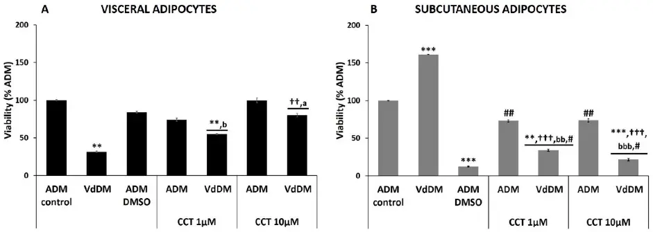
Differentiation of healthy visceral (HV) preadipocytes in VdDM‑conditioned media significantly reduced viability to 31.6 ± 1.0% vs. control (p< 0.01). Co‑treatment with 10 µM CCT restored viability to 80.2 ± 2.4% (p< 0.01 vs. VdDM; ns vs. ADM+CCT 10 µM), whereas 1 µM CCT was ineffective. In contrast, subcutaneous (HS) preadipocyte differentiation in VdDM media increased viability (161.3 ± 0.1%, p< 0.001 vs. control); CCT (1–10 µM) dose‑dependently reduced this aberrant hyper‑viability toward baseline without reaching DMSO toxicity alone (Fig. 1). These findings indicate that a VdDM secretome impairs visceral adipocyte viability while hyper‑stimulating subcutaneous adipogenesis, and that 10 µM crocetin selectively rescues visceral adipocyte viability, supporting a depot‑specific protective role against diabetic adipose dysfunction.
Ask a Question
Write your own review
- You May Also Need
Description: Preadipocytes, found in adipose tissues throughout adulthood, can proliferate and differentiate into mature adipocytes based on energy balance, which in turn increases adipose tissue mass. In vitro ...
Description: 3T3-L1 murine adipocytes have been fundamental in metabolic disease research for over 30 years. Originally derived from Swiss mouse embryo tissue by Dr. Howard Green of Harvard Medical School, the ...
Description: Visceral preadipocytes can be cultured as growing precursor cells or differentiated into adipocytes using medium supplemented with adipogenic and lipogenic hormones. This instruction manual describes ...
Description: Preadipocytes are isolated from omental, mesenteric, or perirenal adipose tissue and are tested for their ability to differentiate in culture to mature adipocytes. Preadipocytes are available ...
Description: Diseased Preadiopcytes are isolated from diabetic type I or type II donors. Further donor information may be available including BMI, medications taken, and disease duration.Preadipocytes are ...
Description: Microvascular endothelial cells line blood vessels and contribute to many biological processes such as angiogenesis, coagulation, trafficking of lymphocytes, and the inflammatory response. ...