
Human Adipose Microvascular Endothelial Cells (HAMEC)
Cat.No.: CSC-7846W
Species: Human
Source: Adipose
Cell Type: Endothelial Cell; Microvascular Cell
- Specification
- Background
- Scientific Data
- Q & A
- Customer Review
Human Adipose Microvascular Endothelial Cells (HAMVECs) are specialized endothelial cells isolated from the microvasculature of human adipose tissue. Given the critical role of adipose tissue as a dynamic endocrine organ, HAMVECs represent a physiologically relevant and high-impact model for studying the interplay between the vascular system, metabolic regulation, and tissue regeneration.
- Tissue-Specific Metabolic Fidelity: Unlike generic macrovascular cells (e.g., HUVECs), HAMVECs retain the unique metabolic and signaling profile of the adipose microenvironment. This makes them the gold standard for research into obesity-related vascular dysfunction, insulin resistance, and type 2 diabetes.
- Robust Angiogenic Potential: HAMVECs exhibit exceptional proliferation and tube-formation capabilities. They are highly responsive to pro-angiogenic factors (e.g., VEGF, bFGF), providing a superior platform for evaluating wound healing, tissue engineering, and anti-angiogenic drug candidates.
- Inflammatory and Adhesion Dynamics: These cells are validated for the expression of key adhesion molecules (ICAM-1, VCAM-1, and E-selectin) and respond dynamically to inflammatory cytokines. They offer a precise model for studying leukocyte recruitment and chronic low-grade inflammation within the fat depot.
- High Purity and Characterization: Our HAMVECs are strictly verified for endothelial markers including CD31 (PECAM-1), vWF, and DiI-Ac-LDL uptake, ensuring they are free from preadipocyte or fibroblast contamination for clean, reproducible experimental results.
By integrating HAMVECs into your research, you gain a human-derived, specialized tool that bridges the gap between systemic metabolic studies and cellular vascular biology.
Isolation of Primary HSaVEC, HITAEC, and HMVEC from Patients Undergoing Coronary Artery Bypass Graft Surgery
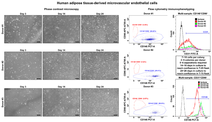
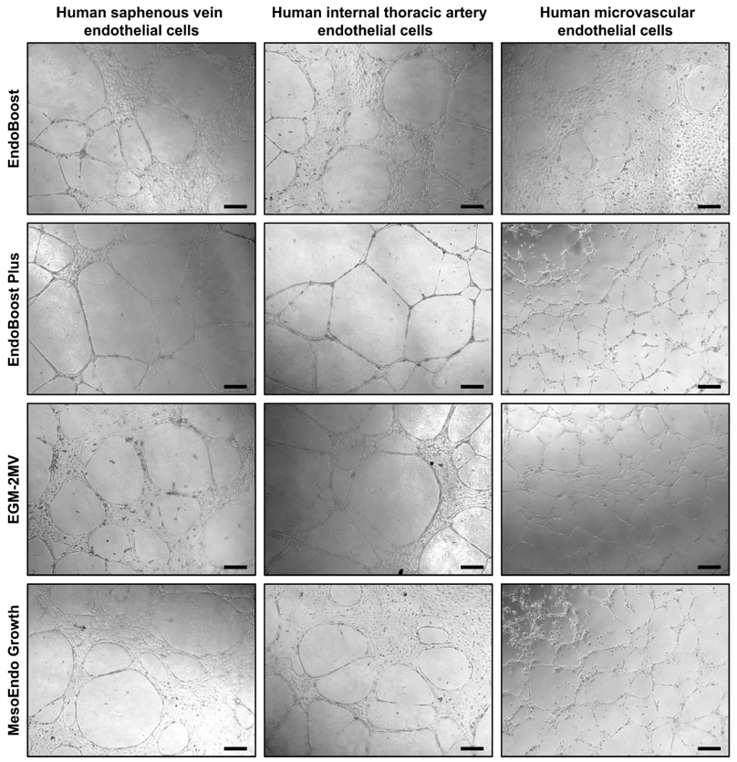
Primary human endothelial cells represent an essential tool to model endothelial dysfunction and to screen interventions for its treatment. Here, we developed a protocol for the synchronous isolation of primary human saphenous vein endothelial cells (HSaVEC), human internal thoracic artery endothelial cells (HITAEC), and human microvascular endothelial cells (HMVEC) from saphenous vein and internal thoracic artery utilized as conduits during coronary artery bypass graft surgery and from subcutaneous adipose tissue excised while providing an access to the heart. Treatment by collagenase type IV and magnetic separation with anti-CD31-antibody-coated beads ensured relatively high efficiency of the isolation (~60% for HSaVEC, ~50% for HITAEC, and ~20% for HMVEC) and high purity (≥99%) of isolated ECs within ~2 weeks (HSaVEC), ~2-3 weeks (HITAEC), and ~3-4 weeks (HMVEC). A colorimetric assay of cell viability and proliferation, as well as real-time bioimpedance monitoring demonstrated high proliferative activity in HSaVEC, HITAEC, and HMVEC, whilst the in vitro tube formation assay indicated their angiogenic potential. The isolation of HSaVEC, HITAEC, and HMVEC from patients undergoing coronary artery bypass graft surgery is a promising option to investigate endothelial heterogeneity, to interrogate endothelial responses to various stresses, and to pinpoint the optimal approaches for restoring endothelial homeostasis, thereby reproducing them within the bedside-to-bench-to-bedside concept.
Studies have shown that angiogenesis precedes adipogenesis, implying that microvascular endothelial cells influence the proliferation of preadipocytes. At the same time, microvascular endothelial cell growth is stimulated by adipocyte secreted VEGG, suggesting a complex paracrine relationship between microvascular endothelial cells and preadipocytes during tissue development.
You will receive Human Adipose Microvascular Endothelial Cells (HAMEC) at passages 2 or 3, depending on cell purity.
It is recommended to use SuperCult® Custom-Made Endothelial Cell Medium (cat# CM-1085X) for the culturing of HAMEC.
Ask a Question
Average Rating: 5.0 | 1 Scientist has reviewed this product
Excellent reproducibility
The cell product has excellent reproducibility.
18 May 2022
Ease of use
After sales services
Value for money
Write your own review
Description: Preadipocytes, found in adipose tissues throughout adulthood, can proliferate and differentiate into mature adipocytes based on energy balance, which in turn increases adipose tissue mass. In vitro ...

Description: 3T3-L1 murine adipocytes have been fundamental in metabolic disease research for over 30 years. Originally derived from Swiss mouse embryo tissue by Dr. Howard Green of Harvard Medical School, the ...
Description: Diseased Preadiopcytes are isolated from diabetic type II donors. Further donor information may be available including BMI, medications taken, and disease duration.Preadipocytes are isolated from ...

Description: Visceral preadipocytes can be cultured as growing precursor cells or differentiated into adipocytes using medium supplemented with adipogenic and lipogenic hormones. This instruction manual describes ...

Description: Preadipocytes are isolated from omental, mesenteric, or perirenal adipose tissue and are tested for their ability to differentiate in culture to mature adipocytes. Preadipocytes are available ...
Description: Diseased Preadiopcytes are isolated from diabetic type I or type II donors. Further donor information may be available including BMI, medications taken, and disease duration.Preadipocytes are ...