
Omental Preadipocytes
Cat.No.: CSC-7657W
Species: Human
Source: Adipose
Cell Type: Preadipocyte
- Specification
- Background
- Scientific Data
- Q & A
- Customer Review
The greater omentum is the largest peritoneal fold within the abdomen and has an immunologic function. The omentum aids in isolating peritoneal infection and absorbing contaminants through the mesothelial stomata. Adipokines and fatty acids from this depot have direct access to the liver through the portal circulation which may lead to hepatic dysfunction.
Omental preadipocytes are primary mesenchymal progenitors isolated from the stromal vascular fraction of the greater omentum, the quintessential visceral adipose depot. In humans, these non-immortalized cells inherently retain the distinct developmental and metabolic identity of intra-abdominal fat, providing a physiologically faithful system that contrasts sharply with subcutaneous preadipocytes.
Their principal advantage as a model lies in replicating the hallmark features of visceral obesity: they display high basal and catecholamine-stimulated lipolytic rates and marked resistance to the anti-lipolytic action of insulin, thereby accurately mirroring the elevated free fatty acid flux that drives hepatic insulin resistance. Concomitantly, they exhibit a pro-inflammatory secretome, constitutively secreting elevated IL-6, TNF-α, and MCP-1 while producing relatively low adiponectin, capturing the chronic low-grade inflammation characteristic of visceral adipose tissue.
Human omental preadipocytes are uniquely sensitive to glucocorticoids due to abundant expression of the glucocorticoid receptor and 11β-hydroxysteroid dehydrogenase type 1, making them an exquisite model for cortisol-induced central adipogenesis. The cells remain amenable to lentiviral gene delivery and siRNA-mediated knockdown and can be co-cultured with macrophages to dissect paracrine inflammatory signaling. Their finite but sufficient proliferative capacity ensures experimental consistency across assays.
Omental Preadipocytes Support Ovarian Cancer Cell Growth and Survival
Ovarian cancer can metastasize to the omentum, which is associated with a complex tumor microenvironment. Omental stromal cells facilitate ovarian cancer colonization by secreting cytokines and growth factors. An improved understanding of the tumor-supportive functions of specific cell populations in the omentum could identify strategies to prevent and treat ovarian cancer metastasis. Here, Waters, Jennifer A., et al. showed that omental preadipocytes enhance the tumor initiation capacity of ovarian cancer cells.
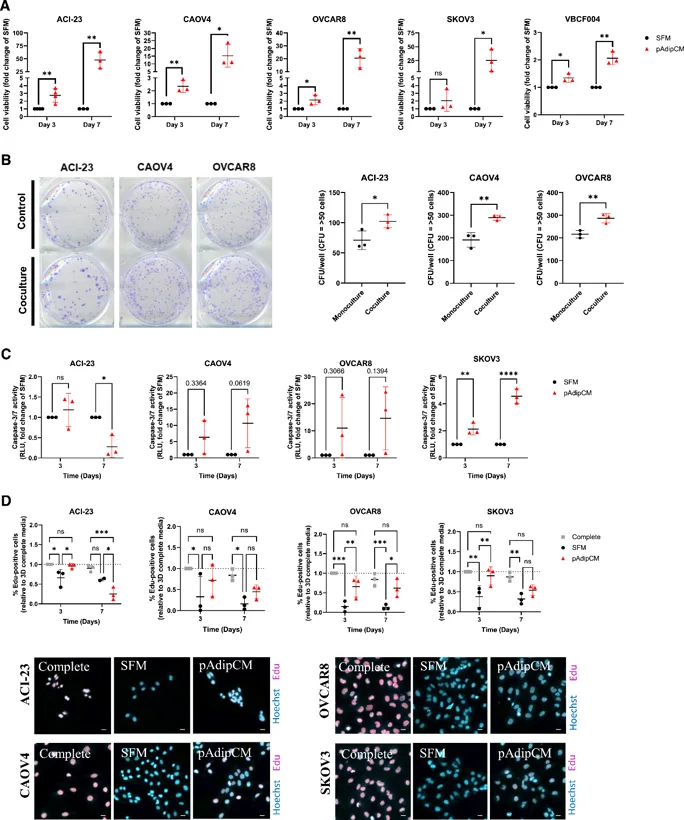
Preadipocytes were cultured in serum-free media (SFM) for 48 hours to generate CM enriched with preadipocyte-secreted factors. At 3 days, CM enhanced cell viability in ACI-23, CAOV4, OVCAR8, SKOV3, and VCBF004, which is an ascites-derived primary cell line. This difference in viability was increased at 7 days and was significant in all cell lines tested (Fig. 1A). Coculture with preadipocytes additionally increased colony formation in all cell lines tested (ACI-23, CAOV4, and OVCAR8; Fig. 1B). Given that increased viability could be due to either reduced apoptosis or increased proliferation, they measured apoptosis via caspase-3/7 activity and proliferation via EdU incorporation either in SFM or CM. In ACI-23 cells, apoptosis decreased in response to CM at 7 days; however, apoptosis was significantly increased in SKOV3 cells (Fig. 1C). A decrease in proliferation was consistent in all cell lines treated with SFM relative to growth in complete media, as expected, and treatment with CM partially rescued this phenotype in all cell lines at Day 3, and all HGSOC cell lines at Day 7 (Fig. 1D).
The isolated omental preadipocytes are plated into the culture ware and treated with our SuperCult® Preadipocyte Differentiation Medium Kit (cat# CM-1145X) to stimulate adipocyte differentiation.
Oil droplets should appear within 4-7 days after differentiation is induced. They look extremely small initially, continue to accumulate and gradually fuse into several big locules.
Adipocytes retain similar morphology and express adipocyte specific genes for at least 4 weeks after induction of differentiation.
The average doubling time is 48-84 hours. However, keep in mind that the replication rate for human preadipocytes varies slightly from patient to patient.
Ask a Question
Average Rating: 5.0 | 1 Scientist has reviewed this product
Happy customer
Happy to be a customer.
24 Apr 2021
Ease of use
After sales services
Value for money
Write your own review
Description: Preadipocytes, found in adipose tissues throughout adulthood, can proliferate and differentiate into mature adipocytes based on energy balance, which in turn increases adipose tissue mass. In vitro ...
Description: 3T3-L1 murine adipocytes have been fundamental in metabolic disease research for over 30 years. Originally derived from Swiss mouse embryo tissue by Dr. Howard Green of Harvard Medical School, the ...
Description: Diseased Preadiopcytes are isolated from diabetic type II donors. Further donor information may be available including BMI, medications taken, and disease duration.Preadipocytes are isolated from ...
Description: Visceral preadipocytes can be cultured as growing precursor cells or differentiated into adipocytes using medium supplemented with adipogenic and lipogenic hormones. This instruction manual describes ...
Description: Preadipocytes are isolated from omental, mesenteric, or perirenal adipose tissue and are tested for their ability to differentiate in culture to mature adipocytes. Preadipocytes are available ...
Description: Diseased Preadiopcytes are isolated from diabetic type I or type II donors. Further donor information may be available including BMI, medications taken, and disease duration.Preadipocytes are ...