
Human Choroid Plexus Endothelial Cells (HCPEC)
Cat.No.: CSC-7808W
Species: Human
Source: Brain
Cell Type: Endothelial Cell
- Specification
- Background
- Scientific Data
- Q & A
- Customer Review
Human Choroid Plexus Endothelial Cells (HCPEC) are primary endothelial cells isolated from the vascular compartment of the human choroid plexus, a highly specialized tissue situated within the brain ventricles. HCPECs are characterized by fenestrated endothelium and play a crucial role in maintaining the blood-cerebrospinal fluid barrier (BCSFB), while brain microvascular endothelial cells constitute the blood-brain barrier (BBB). These cells play an important part in the maintenance of cerebrospinal fluid balance, molecular transport, immunological surveillance and neurovascular communication in the central nervous system (CNS).
HCPECs display classical endothelial markers such as CD31, VE-cadherin and von Willebrand factor (vWF) as well as functional characteristics of the choroid plexus vasculature. They provide a useful in vitro model to study endothelial transport processes, leukocyte trafficking, inflammatory responses and interactions between the vascular and epithelial compartments of the choroid plexus. Endothelial cells from the human choroid plexus are frequently used in research of blood-CSF barrier physiology, neuroinflammation, CNS infections, neurodegenerative disorders and medication transport across brain barriers. In co-culture with choroid plexus epithelial cells, HCPECs can be used to develop physiologically appropriate BCSFB models closely resembling the in vivo milieu. These models are powerful instruments for assessing barrier integrity, permeability, therapeutic delivery, and pathological processes involved in neurological illnesses, which makes HCPECs extremely relevant for neuroscience research, translational studies, and CNS drug development.
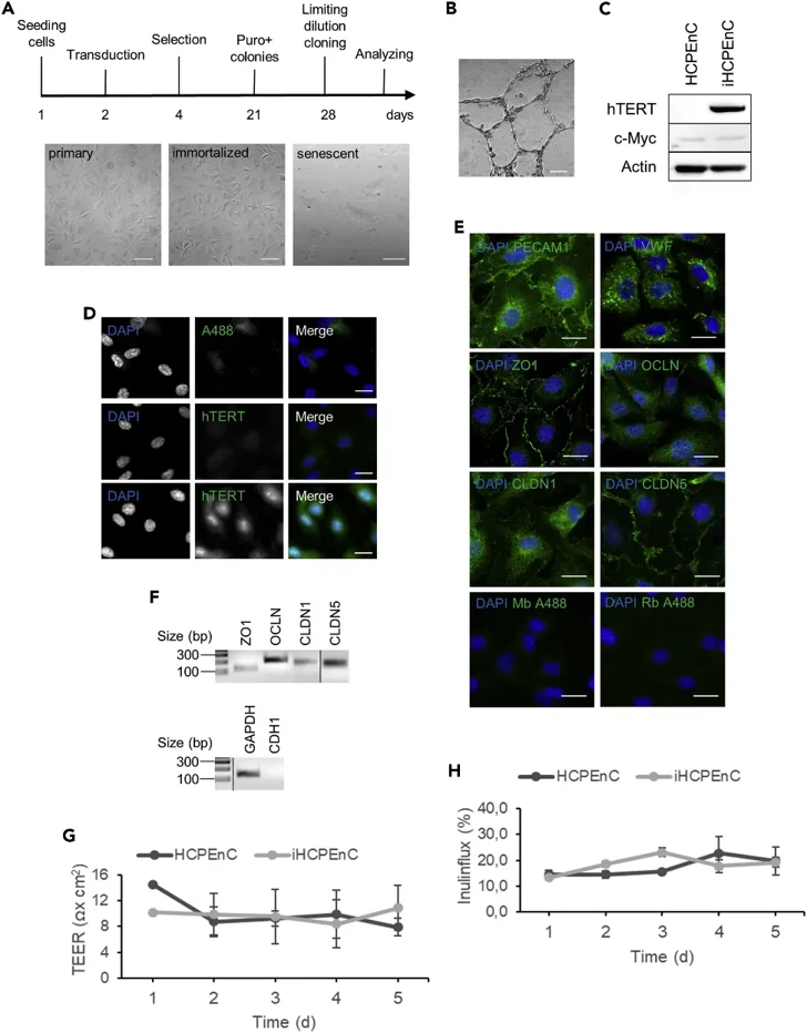
Transcriptomic Stability of Immortalized Human Choroid Plexus Endothelial Cells
The choroid plexus (CP) forms the blood–cerebrospinal fluid barrier (BCSFB). To enable sustained study, Denzer et al. immortalized primary human CP endothelial cells (HCPEnCs) via hTERT expression. Here, they compared the transcriptomes of primary HCPEnCs (passage 6; p6) with low (p20) and high (p50) passage immortalized cells (iHCPEnCs).
Comparative analysis of 26,586 transcripts revealed high overall concordance. Filtering for significance (log₂FC > 1.0, p < 0.05, FDR < 0.05) identified 2,899 (p20_vs_p6), 3,038 (p50_vs_p6), and 4,791 (p50_vs_p20) differentially expressed genes (DEGs) (Fig. 1a). Overlap analysis clarified the relationship between these DEGs (Fig. 1b). Comparing p20_vs_p6 and p50_vs_p6 identified 1,806 genes unique to p20, 1,945 unique to p50, and 1,093 shared DEGs between iHCPEnCs and primary cells. Contrasting these with p50_vs_p20 revealed that 769 genes were altered specifically relative to the primary state but not between immortalized passages. Conversely, 4,467 genes differed between p50 and p20 without significant change relative to primary cells. Only 324 genes overlapped between these categories.
These data demonstrate that while iHCPEnCs maintain high transcriptomic fidelity to primary cells, distinct gene sets are modulated at low versus high passage, informing their appropriate use in barrier research.
Ask a Question
Write your own review

Description: Creative Bioarray's Human Monocyte-Derived Dendritic Cells provide an ideal culture model for the study of the initiation of immune responses, delivery of antigens to dendritic cells, cytokine ...

Description: GFP Expressing Human Astrocytes (GFP-HAs) provided by Creative Bioarray are puromycin-selected from primary human astrocytes infected with lentivirus expressing GFP. GFP-HAs are negative for ...
Description: Human Neurons-hippocampal (HN-h) from Creative Bioarray are isolated from hippocampal tissue of the brain. HHN are cryopreserved at primary cultures and delivered frozen. Each vial contains >1 x 10^6 ...
Description: Human Astrocytes-midbrain (HA-mb) from Creative Bioarray are isolated from human midbrain (mesencephalon). HA-mb are cryopreserved at passage one and delivered frozen. Each vial contains >5 x 10^5 ...
Description: Human Choroid Plexus Fibroblasts (HCPF) from Creative Bioarray are isolated from human choroid pluxus. HCPF are cryopreserved at primary culture and delivered frozen. Each vial contains >5 x 10^5 ...
Description: HN are isolated from the human brain. HN are cryopreserved at primary cultures and delivered frozen. Each vial contains >1 x 10^6 cells in 1 ml volume. HN are characterized by immunofluorescent ...