
Human Mammary Microvascular Endothelial Cells
Cat.No.: CSC-C8612W
Species: Human
Source: Breast
Cell Type: Endothelial Cell; Microvascular Cell
- Specification
- Background
- Scientific Data
- Q & A
- Customer Review
Human Mammary Microvascular Endothelial Cells (HMEC) are primary cells obtained from the microvascular endothelial layer of mammary gland. They constitute the inner lining of tiny blood arteries in breast tissue and are important for vascular homeostasis, nutrition exchange, and immune cell trafficking. In culture, HMEC form a typical cobblestone-like monolayer at confluence and require a specialized basal media supplemented with growth factors (e.g., VEGF) and serum to maintain their quiescent, differentiated phenotype. Standard cultivation conditions are 37 °C and 5 % CO2.
HMEC are a physiologically appropriate model for the study of mammary gland angiogenesis, vascular permeability and tumor microenvironment interactions. HMEC enable extravasation and spread of cancer cells in breast cancer. They are also utilized to study endothelial responses to hormones, cytokines and anti-angiogenic treatments, giving us insight into normal breast biology and pathological vascular remodeling.
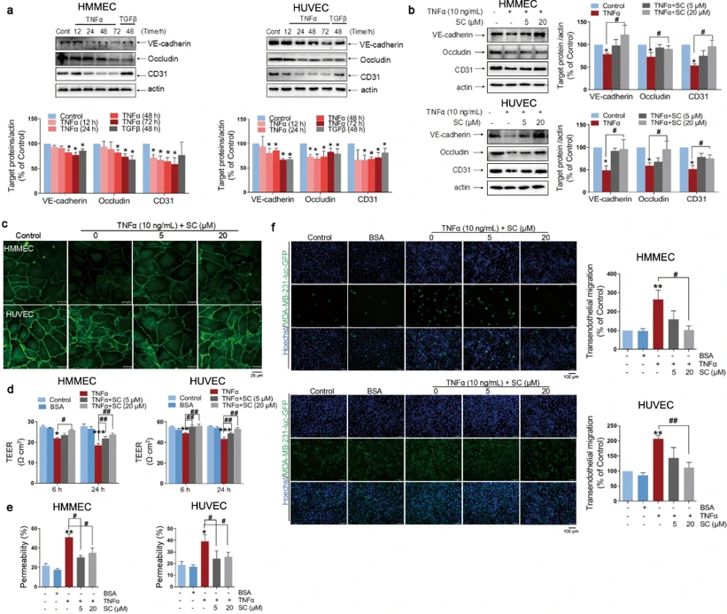
SC Rescued TNFΑ-Induced Endothelial Barrier Dysfunction and Transendothelial Migration in TNBC In Vitro
Triple-negative breast cancer (TNBC) metastasis is exacerbated by tumor-associated vascular abnormalities. While Scutellarin (SC) is known for cardiovascular benefits, its role in mitigating TNBC metastasis via vascular protection is unknown. Mei's team investigated SC's ability to preserve endothelial barrier integrity against inflammatory insults.
TNFα and TGFβ disrupted endothelial tight junctions, decreasing VE-cadherin, occludin, and CD31 expression in HMMECs and HUVECs (Fig. 1a). SC treatment (5-20 µM) reversed this effect, restoring the expression of these junctional proteins (Fig. 1b, c). Functionally, SC rescued the TNFα-induced drop in transepithelial electrical resistance (TEER) (Fig. 1d) and attenuated FITC-dextran leakage (Fig.1e). Crucially, SC suppressed the transendothelial migration of MDA-MB-231 and 4T1 breast cancer cells across TNFα-stimulated endothelial monolayers (Fig. 1f). These data demonstrate that SC fortifies the endothelial barrier, thereby inhibiting tumor cell extravasation.
Ask a Question
Write your own review
Description: Human Thymus Fibroblasts are isolated from normal human thymus tissue.
Description: Creative Bioarray's normal Human Mammary Epithelial Cells, when grown in Creative Bioarray's LIMam Medium, provide an ideal serum-free culture model for many areas of research. Common uses of HMEC ...

Description: Human Breast Tumor-Associated Endothelial Cells from Creative Bioarray are isolated from human breast tumor tissue. Human Breast Tumor-Associated Endothelial Cells are grown in T25 tissue culture ...
Description: Human Breast Carcinoma Epithelial Cells from Creative Bioarray are isolated from human breast tumor tissue. Human Breast Carcinoma Epithelial Cells are grown in T25 tissue culture flasks pre-coated ...
Description: Human Mammary Epithelial Cells (HMECs) produced at Creative Bioarray are isolated from human mammary tissue. The cells are cryopreserved at passage 2 (P2) and delivered frozen. Each vial contains ...
Description: Recent research indicates cancer associated fibroblasts (CAFs) significant involvement in crucial aspects of epithelial solid tumor biology, specifically neoplastic progression, tumor growth, ...