
Human Breast Tumor-Associated Endothelial Cells
Cat.No.: CSC-C8582W
Species: Human
Source: Breast
Cell Type: Endothelial Cell
- Specification
- Background
- Scientific Data
- Q & A
- Customer Review
Human Breast Tumor Associated Endothelial Cells (HBTAECs) are primary endothelial cells isolated from the vasculature of human breast tumor tissues. HBTAECs are not similar to the normal endothelial cells, but they show the specific biological and functional features of the tumor microenvironment, such as increased angiogenesis, abnormal vascular organization and altered gene expression profiles. These cells usually express canonical endothelial markers such as CD31, VE-cadherin and von Willebrand factor (vWF) but also demonstrate increased expression of pro-angiogenic factors including VEGFR2 and heightened sensitivity to tumor-derived signals. HBTAECs were more proliferative, migratory and tube-forming than endothelial cells derived from normal tissues adjacent to the tumors. They also exhibit unique metabolic adaptations and increased resistance to anti-angiogenic therapies, which makes them extremely relevant to the study of therapeutic resistance. Importantly, these cells actively cross-talk with cancer cells, immune cells and stromal components, leading to tumor progression, metastasis and immune modulation.
HBTAECs, due to their tumor-specific phenotype, represent a physiologically relevant in vitro model for investigating breast cancer angiogenesis, vascular permeability and drug delivery mechanisms. They are widely used in anti-angiogenic drug screening, nanomedicine targeting studies and tumor microenvironment research. Overall, HBTAECs are useful platform to understand the intricacies of tumor vasculature and develop more effective targeted cancer therapies.
Tumor-Associated Endothelial Cells Favor Interactions with Administered Nanoparticles
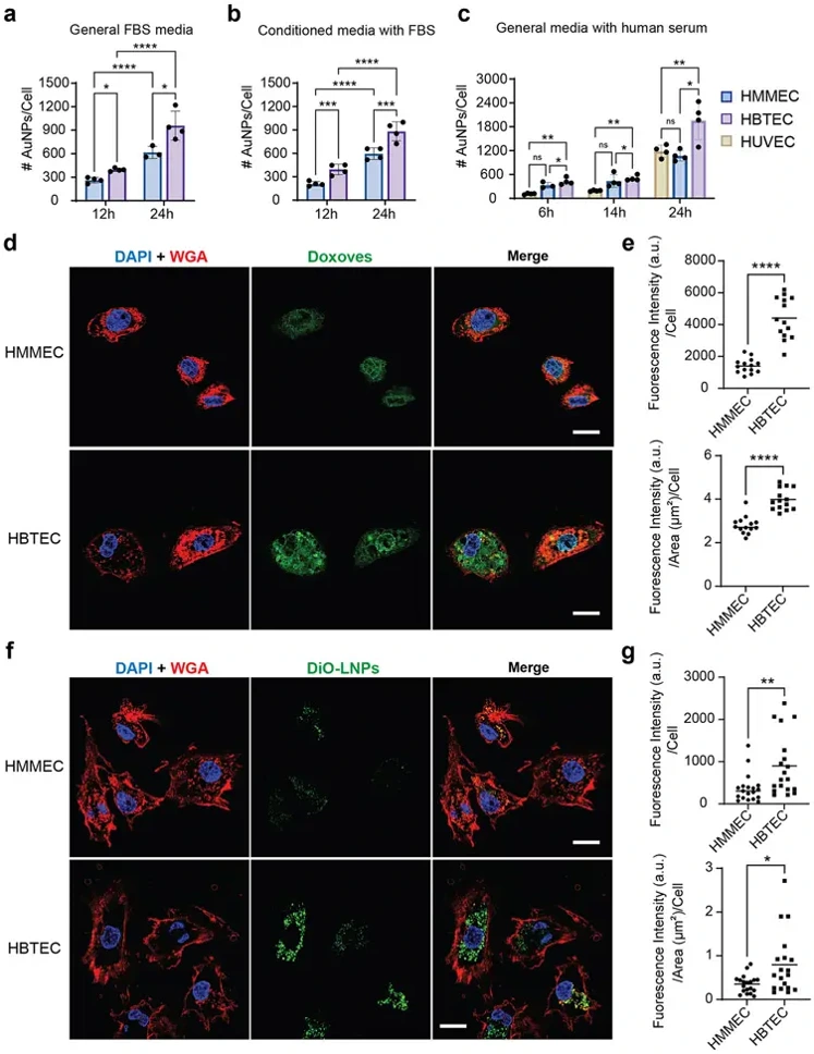
Cancer nanomedicines rely on endothelial cells for tumor delivery, but their translational relevance in humans is unclear. Using primary human breast cancer endothelial cells (HBTECs), Wang et al. quantified the differential interactions of normal versus tumor-associated endothelial cells with three clinically relevant nanomedicines: PEGylated gold nanoparticles, liposomal doxorubicin, and LNPs.
Across all tested conditions-including standard media, conditioned media, and human serum-HBTECs exhibited significantly stronger interactions with gold nanoparticles than normal controls (HMMECs, HUVECs), reaching up to ~1.9-fold higher uptake (Fig. 1a-c). No significant difference was observed between general and conditioned media.
Similarly, confocal microscopy revealed greater intracellular fluorescence in HBTECs following incubation with liposomal doxorubicin or DiO-LNPs (Fig. 1d, f). Quantitative image analysis confirmed higher uptake per cell and per cell area in HBTECs (Fig. 1e, g).
These findings were consistent across nanoparticles with varying sizes (~70-90 nm) and stiffness. Collectively, our data demonstrate that tumor-associated endothelial cells exhibit enhanced interactions with diverse nanomedicines compared to normal endothelial cells.
Ask a Question
Write your own review
Description: Human Thymus Fibroblasts are isolated from normal human thymus tissue.

Description: Human Mammary Microvascular Endothelial Cells from Creative Bioarray are isolated from human breast tissue. Human Mammary Microvascular Endothelial Cells are grown in T25 tissue culture flasks ...

Description: Creative Bioarray's normal Human Mammary Epithelial Cells, when grown in Creative Bioarray's LIMam Medium, provide an ideal serum-free culture model for many areas of research. Common uses of HMEC ...
Description: Human Breast Carcinoma Epithelial Cells from Creative Bioarray are isolated from human breast tumor tissue. Human Breast Carcinoma Epithelial Cells are grown in T25 tissue culture flasks pre-coated ...
Description: Human Mammary Epithelial Cells (HMECs) produced at Creative Bioarray are isolated from human mammary tissue. The cells are cryopreserved at passage 2 (P2) and delivered frozen. Each vial contains ...
Description: Recent research indicates cancer associated fibroblasts (CAFs) significant involvement in crucial aspects of epithelial solid tumor biology, specifically neoplastic progression, tumor growth, ...