
Human Breast Cancer Associated Fibroblasts
Cat.No.: CSC-C30028J
Species: Human
Source: Breast
Cell Type: Fibroblast
- Specification
- Background
- Scientific Data
- Q & A
- Customer Review
Within the breast tumor microenvironment (TME), cancer-associated fibroblasts (CAFs) constitute the most abundant stromal cell population and serve as the primary architects of the tumor stroma. These specialized mesenchymal cells are not passive bystanders but actively orchestrate tumor progression through extracellular matrix (ECM) remodeling, secretion of bioactive factors, and dynamic interactions with neighboring cells.
CAFs in breast cancer exhibit remarkable functional and phenotypic heterogeneity, originating from diverse cellular precursors including resident fibroblasts, mesenchymal stem cells, pericytes, and even cancer cells undergoing epithelial-mesenchymal transition. High-resolution single-cell RNA sequencing and spatial transcriptomics have identified multiple distinct CAF subtypes, including inflammatory CAFs (iCAFs), myofibroblastic CAFs (myCAFs), matrix CAFs (mCAFs), antigen-presenting CAFs (apCAFs), and proliferative CAFs. These subpopulations exhibit distinct transcriptional programs, spatial distributions, and functional capacities. For instance, myCAFs form a physical barrier around the tumor core that impedes immune cell infiltration, whereas iCAFs co-localize with immune cells in peripheral niches.
The principal advantage of human breast CAFs lies in their utility as a physiologically relevant in vitro model system. These cells are extensively employed in co-culture systems with breast cancer cell lines and patient-derived organoids (PDOs) to recapitulate tumor-stroma interactions, study CAF-mediated therapy resistance, and evaluate therapeutic interventions. Furthermore, CAF-related gene signatures have demonstrated robust prognostic value, effectively stratifying patient survival outcomes and predicting treatment response. The strong correlation between specific CAF subsets and clinical outcomes highlights their potential as both predictive biomarkers and therapeutic targets.
Targeting MCL-1 in bCAFs Enhances Endothelial Cell Tubulogenesis and Angiogenesis
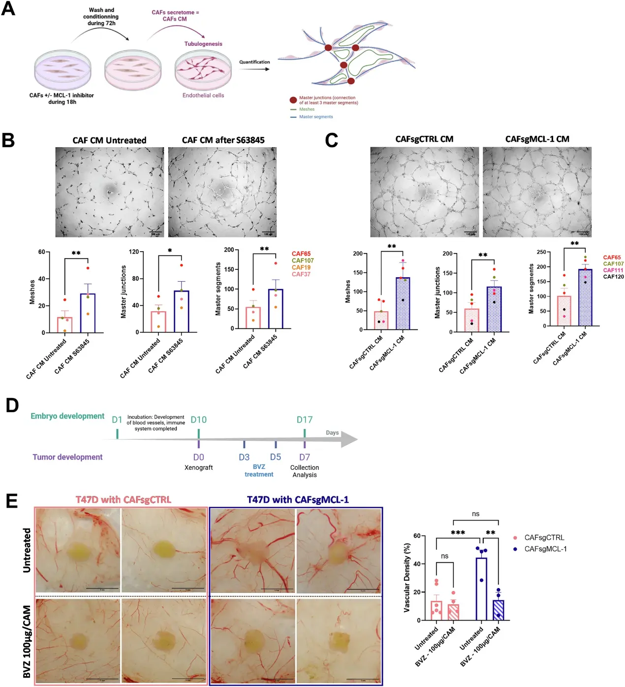
To evaluate the impact of anti-apoptotic protein MCL-1 in breast cancer-associated fibroblasts (bCAFs) on angiogenesis, we generated conditioned media from bCAFs pre-treated with S63845 (CAF CM after S63845) or not (CAF CM Untreated) for 18 h or from bCAFs silenced for MCL-1 (CAFsgMCL-1 CM) or not (CAFsgCTRL CM). We evaluated the capacities of endothelial cells to organize pseudo-like vessels in vitro with tubulogenesis assay in response to bCAFs secretome (Fig. 1A). Conditioned media from bCAFs pre-treated with S63845 significantly promoted tubulogenesis by inducing more master junctions, master segments, and meshes in comparison with conditioned media from untreated bCAFs (Fig. 1B). In parallel, conditioned media from bCAFs silenced for MCL-1 (CAFsgMCL-1 CM) also significantly enhanced the endothelial tubulogenesis in comparison with conditioned media from the control bCAFs (CAFsgCTRL CM) (Fig. 1C). In the chicken chorioallantoic membrane CAM model, we performed xenografts of tumor models composed of T47D luminal breast cancer cells mixed with primary bCAFs genetically modified for MCL-1 (CAFsgMCL-1) or not (CAFsgCTRL) (Fig. 1D). one week after xenografts on CAM, we observed that the peritumoral vascular density is significantly increased around the tumors composed of bCAFsgMCL-1 compared to bCAFsgCTRL (Fig. 1E). Furthermore, these effects are annihilated with VEGF inhibitor treatment Bevacizumab (BVZ) (Fig. 1E). These results highlight the strong involvement of MCL-1 in preventing VEGF-A dependant pro-angiogenic effect of bCAFs.
Ask a Question
Write your own review
Description: Human Thymus Fibroblasts are isolated from normal human thymus tissue.
Description: Human Mammary Microvascular Endothelial Cells from Creative Bioarray are isolated from human breast tissue. Human Mammary Microvascular Endothelial Cells are grown in T25 tissue culture flasks ...
Description: Creative Bioarray's normal Human Mammary Epithelial Cells, when grown in Creative Bioarray's LIMam Medium, provide an ideal serum-free culture model for many areas of research. Common uses of HMEC ...
Description: Human Breast Tumor-Associated Endothelial Cells from Creative Bioarray are isolated from human breast tumor tissue. Human Breast Tumor-Associated Endothelial Cells are grown in T25 tissue culture ...
Description: Human Breast Carcinoma Epithelial Cells from Creative Bioarray are isolated from human breast tumor tissue. Human Breast Carcinoma Epithelial Cells are grown in T25 tissue culture flasks pre-coated ...
Description: Human Mammary Epithelial Cells (HMECs) produced at Creative Bioarray are isolated from human mammary tissue. The cells are cryopreserved at passage 2 (P2) and delivered frozen. Each vial contains ...