
Rat Submandibular Gland Epithelial Cells
Cat.No.: CSC-C5035S
Species: Rat
Source: Salivary Gland
Cell Type: Epithelial Cell
- Specification
- Background
- Scientific Data
- Q & A
- Customer Review
Rat Submandibular Gland Epithelial Cells (rSGECs) from Creative Bioarray are isolated from the rat submandibular gland tissue. The method we use to isolate rSGECs was developed based on a combination of established and our proprietary methods. The rSGECs are characterized by immunofluorescence with antibodies specific to Cytokeratin-8 (CK-8). Each vial contains 0.5x10^6 cells per ml and is delivered frozen.
Rat Submandibular Gland Epithelial Cells are primary epithelial cells derived from the submandibular glands of healthy rats. The submandibular gland is one of the major salivary glands and is involved in saliva production, oral lubrication, digestion, and the maintenance of oral homeostasis. Epithelial cells are the main functional cells in the gland and they are organized into acinar and ductal structures that secrete, modify and transport saliva. These cells therefore offer a physiologically relevant in vitro model for the investigation of salivary gland function and epithelial biology.
Rat submandibular gland epithelial cells have a polygonal cobblestone-like morphology and grow as an adherent monolayer in culture. They express epithelial markers cytokeratins and epithelial cell adhesion molecules. They retain characteristics associated with salivary gland epithelial function. These cells respond to neurotransmitters, growth factors, inflammatory mediators, and extracellular matrix cues that regulate epithelial proliferation, differentiation, secretion, and tissue remodeling.
Rat Submandibular Gland Epithelial Cells are routinely used in studies of salivary gland physiology, epithelial regeneration and glandular injury. These are useful experimental platforms to study mechanisms of salivary secretion, epithelial barrier function, oxidative stress, inflammatory responses, cell-matrix interactions. These cells are also used to study radiation-induced damage of salivary glands, autoimmune diseases of the salivary glands, tissue engineering, regenerative medicine, and testing of therapeutic agents to maintain or restore salivary gland function.
Acetylcholine Does Not Affect Branching or Proliferation but Is Present During Submandibular Gland Myoepithelial Differentiation
Myoepithelial cells (MECs) contract to expel saliva under parasympathetic control, yet the molecular signals driving SG epithelial cells to differentiate into MECs adjacent to parasympathetic nerves remain unclear. Shindo et al. used an ex vivo embryonic submandibular gland (SMG) organ culture model with carbachol (CCh), a cholinergic agonist, to test whether acetylcholine signaling influences SMG development.
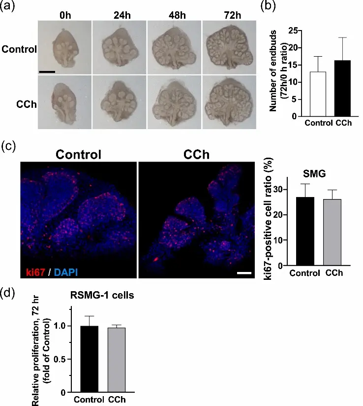
E13.5 SMGs cultured 72 h with CCh showed no significant differences in branching morphogenesis or end‑bud number compared to control (Fig. 1a, b). Similarly, tetrodotoxin (TTX, 10 µM), a Na⁺‑channel blocker, did not alter branching, indicating TTX‑sensitive neuronal activity is not required for early SMG morphogenesis.
Proliferation was assessed by Ki67 immunostaining in situand CCK‑8 assay on the rat SG epithelial cell line RSMG‑1. The proportion of Ki67⁺ epithelial cells in end‑buds was unchanged by CCh treatment (Fig. 1c), and RSMG‑1 cell proliferation was likewise unaffected (Fig. 1d).
These results demonstrate that cholinergic agonist exposure does not alter SMG branching morphogenesis or epithelial cell proliferation, establishing a baseline for investigating acetylcholine’s specific role in inducing myoepithelial cell differentiation near parasympathetic nerve terminals.
Ask a Question
Write your own review
- You May Also Need
Description: The thoracic aorta is located in the chest cavity and gives off arteries that branch to the esophagus, pericardium, lungs, and trachea. The thoracic aorta can be subdivided into the ascending aorta, ...
Description: Rat Podocytes are isolated from normal rat kidney. The cells are characterized by immunofluorescence with antibodies specific to podocin, Ang1, Nephrin, ACTN4, NPHS2. T25 flasks is required for cell ...
Description: Rat Bronchial Smooth Muscle Cells are isolated from normal rat bronchi tissue. Rat Bronchial Smooth Muscle Cells are characterized by immunofluorescence with antibodies specific to alpha-actin. T25 ...
Description: Guinea Pig Endothelial Cells from Creative Bioarray are isolated from guinea pig tissue. Prior to shipping, cells at passage 2 are detached from flasks and immediately cryopreserved in vials. Each ...
Description: Rat Lung Epithelial Cells are isolated from normal rat lung tissue. The cells are characterized by immunofluorescence with antibodies specific to CK-18, CK-19. T25 flasks is required for cell ...
Description: Rat Vein Endothelial Cells from Creative Bioarray are isolated from inferior vena cava tissue of 6-8 week old laboratory Sprague-Dawley rat. Rat Vein Endothelial Cells are grown in T75 tissue culture ...