
Rat Podocytes
Cat.No.: CSC-C9378W
Species: Rat
Source: Kidney
Morphology: Epithelial-like
Cell Type: Podocyte
- Specification
- Background
- Scientific Data
- Q & A
- Customer Review
Rat podocytes, a primary glomerular cell type of rat kidney podocyte lines, have been used extensively as a model for kidney function and pathology. In the glomerulus, these visceral epithelial cells attach to the basement membrane surrounding the capillaries via branching protrusions in a sophisticated tertiary "foot" process network. These processes bundle together to make a slit diaphragm, which prevents the accumulation of proteins and other large molecules in the urine, keeping the structure of the glomerular filtration barrier intact.
The cytoskeleton of podocytes is rich in actin, allowing them to dynamically remodel and adapt to pressure changes. Their complex shape and molecular dynamics keep the flux of substances between blood and urine exactly under control, maintaining the internal environment. In immunofluorescence testing, podocyte markers can be identified including podocin, angiotensin 1, nephrin, actin 4, and NPHS2. In clinical research, rat podocyte lines play an essential role in the study of glomerular disease, especially diabetic nephropathy and membranous nephropathy. The lineages of in vitro cultured podocytes offer a robust model for understanding podocyte biology, molecular biology and drug discovery. They are also critical for examining cell signaling pathways, mechanisms of podocyte damage and therapeutic strategies, helping to uncover the underlying pathology of renal disease and experimentally supporting new therapies.
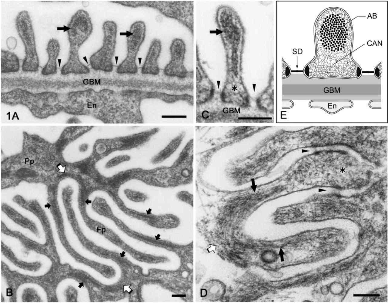 Fig. 1. Electron micrographs of rat podocyte foot processes. (a) Perpendicular sections. (b, d) Horizontal sections. (c) Cross-sections (Ichimura K, Kurihara H, et al., 2003).
Moringa oleifera Leaf Extract Enhances the Viability and Inhibits Ferroptosis in High Glucose-induced Rat Podocytes
Diabetic nephropathy (DN) is a severe complication in patients with diabetes mellitus, which is mainly characterized by glomerular podocyte injury. Ferroptosis is crucial in the pathogenesis of DN. Moringa oleifera leaf extract (MOLE) has attracted attention due to its multiple pharmacological activities, but it is unclear whether it can delay the progression of DN by inhibiting ferroptosis.
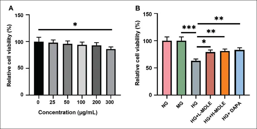
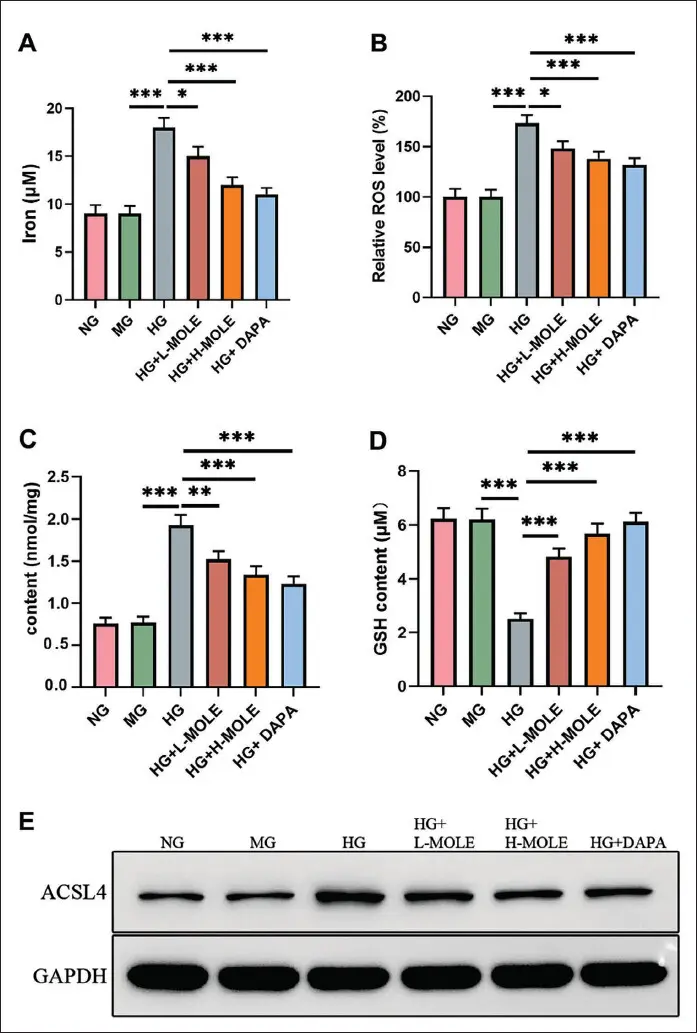
DN model cells were established using rat podocytes induced by high glucose (HG); the cells were processed with MOLE, dapagliflozin (DAPA, positive control), and acyl-CoA synthetase long-chain family member 4 (ACSL4) overexpression plasmids. Cell viability, Fe2+ concentration, reactive oxygen species (ROS), malondialdehyde (MDA), and glutathione (GSH) levels were monitored.
MOLE treatment markedly enhanced HG-induced rat podocyte viability, decreased intracellular Fe2+ concentration, ROS level, MDA concentration, and elevated GSH concentration. ACSL4 overexpression reversed the protective effect of MOLE on podocytes. This finding provides a theoretical basis for further developing MOLE-based therapeutic agents for DN treatment.
Ask a Question
Write your own review
Description: Characterization: 1. Cell Activity; Recovery Viability ≥ 80%. 2. Cell Purity; - GFAP-positive cell percentage ≥ 70%. - β-tubulin III- or doublecortin- (Dcx-) positive cell percentage ≤ 10%. - ...
Description: Chondrocytes are the cells responsible for cartilage formation, and they are crucial for the process of endochondral ossification, which is useful for bone development. Ear chondrocytes represent the ...
Description: The thoracic aorta is located in the chest cavity and gives off arteries that branch to the esophagus, pericardium, lungs, and trachea. The thoracic aorta can be subdivided into the ascending aorta, ...
Description: Rat Bone Marrow Neutrophils are derived from the tibias and femurs of week-5 Sprague–Dawley Rats. Rat Bone Marrow Neutrophils are immediately cryo-preserved or fresh cells in suspension after ...

Description: Rat Bone Marrow Macrophages are derived from the tibias and femurs of 5 weeks old Sprague–Dawley Rats. Rat Bone Marrow Macrophages are grown in 100 mm treated tissue culture dish and incubated in ...
Description: The abdominal aorta is the largest artery in the abdominal cavity. As part of the aorta, it is a direct continuation of the descending aorta. There are three layers to the aortic wall, the tunica ...