
Rat Skeletal Muscle Fibroblasts
Cat.No.: CSC-C5121S
Species: Rat
Source: Skeletal Muscle
Cell Type: Fibroblast
- Specification
- Background
- Scientific Data
- Q & A
- Customer Review
Rat Skeletal Muscle Fibroblasts (rSMFs) from Creative Bioarray are isolated from the rat leg muscle tissue. The method we use to isolate rSMFs was developed based on a combination of established and our proprietary methods. The rSMFs are characterized by immunofluorescence with antibodies specific to vimentin. Each vial contains 0.5x10^6 cells per ml and is delivered frozen.
Rat Skeletal Muscle Fibroblasts are main stromal cells derived from rat skeletal muscle tissue. These cells are key components of the skeletal muscle milieu and play a crucial role in the maintenance, remodeling and repair of muscle tissue through the synthesis and organization of extracellular matrix (ECM) proteins. These cells are involved in tissue architecture and interact with muscle fibres, satellite cells, immune cells and vascular cells under physiological settings.
Rat Skeletal Muscle Fibroblasts are frequently utilized to research extracellular matrix dynamics, fibroblast activation, tissue remodeling, and cellular interactions in skeletal muscle. These cells may become activated upon injury or pathological stimulation and contribute to matrix protein production and modulation of tissue repair processes. They are therefore often used in studies of muscle fibrosis, regenerative responses and signaling pathways in stromal cell activity.
These cells are also a helpful in vitro model for examining factors that modulate fibroblast behavior, including cytokines, growth hormones, mechanical stimulation, and pharmacological agents. More recently, rat skeletal muscle fibroblasts have been used to study TGF-β-mediated fibrogenic signaling, extracellular matrix remodeling, fibroblast-myocyte interactions, and age-related alterations in skeletal muscle tissue.
Calcitriol Suppresses Skeletal Muscle Fibroblast Proliferation via Vitamin D Receptor Signaling
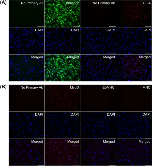
Fibroblasts are central mediators of fibrogenesis following skeletal muscle injury, contributing to excessive scarring and impaired functional recovery. Although vitamin D receptor (VDR) expression is elevated in regenerating muscle, the direct effects of calcitriol [1α,25(OH)₂D₃] on muscle fibroblasts remain unclear. Srikuea et al. characterized primary fibroblasts isolated from the skeletal muscle of 1-month-old male C57BL/6 mice. Cells were confirmed as fibroblastic based on vimentin⁺/TCF-4⁺ immunostaining and the absence of myogenic differentiation markers (EbMHC and MHC) under differentiation conditions, verifying a non-myogenic, fibroblast-dominant population (Fig. 1A, B).
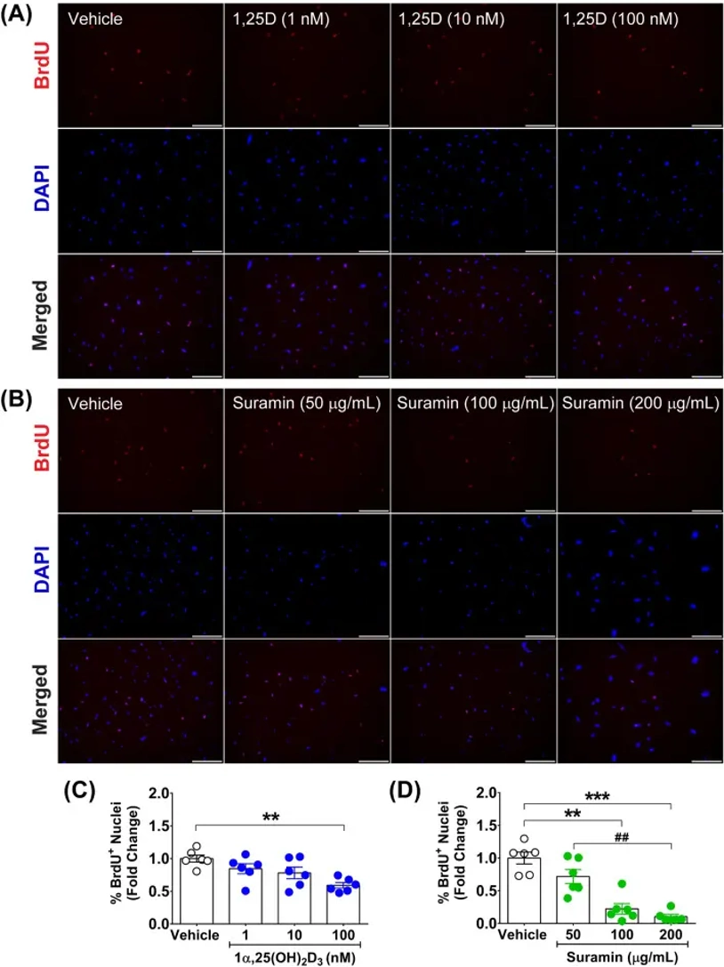
To assess antiproliferative effects, cells were treated daily for 48 hours with calcitriol (1, 10, 100 nM) or the antifibrotic agent suramin (50, 100, 200 µg/mL). BrdU pulse-labeling demonstrated a significant reduction in S-phase entry following both treatments (Fig. 2A, B). Quantitative analysis revealed that 100 nM calcitriol suppressed proliferation to 0.59 ± 0.04-fold of vehicle control (p< 0.01) (Fig. 2C). In contrast, suramin exhibited stronger potency, reducing proliferation to 0.22 ± 0.08-fold at 100 µg/mL (p< 0.01) and 0.10 ± 0.03-fold at 200 µg/mL (p< 0.001) (Fig. 2D).
These findings demonstrate that calcitriol directly inhibits skeletal muscle fibroblast proliferation in vitro. While effective, its antiproliferative capacity is less potent than that of suramin at efficacious concentrations, highlighting its potential as a moderate modulator of fibroproliferative processes in muscle injury.
Ask a Question
Write your own review
- You May Also Need
Description: The thoracic aorta is located in the chest cavity and gives off arteries that branch to the esophagus, pericardium, lungs, and trachea. The thoracic aorta can be subdivided into the ascending aorta, ...
Description: Rat Podocytes are isolated from normal rat kidney. The cells are characterized by immunofluorescence with antibodies specific to podocin, Ang1, Nephrin, ACTN4, NPHS2. T25 flasks is required for cell ...
Description: Rat Bronchial Smooth Muscle Cells are isolated from normal rat bronchi tissue. Rat Bronchial Smooth Muscle Cells are characterized by immunofluorescence with antibodies specific to alpha-actin. T25 ...
Description: Guinea Pig Endothelial Cells from Creative Bioarray are isolated from guinea pig tissue. Prior to shipping, cells at passage 2 are detached from flasks and immediately cryopreserved in vials. Each ...
Description: Rat Lung Epithelial Cells are isolated from normal rat lung tissue. The cells are characterized by immunofluorescence with antibodies specific to CK-18, CK-19. T25 flasks is required for cell ...
Description: Rat Vein Endothelial Cells from Creative Bioarray are isolated from inferior vena cava tissue of 6-8 week old laboratory Sprague-Dawley rat. Rat Vein Endothelial Cells are grown in T75 tissue culture ...